Search results (431 results)
-
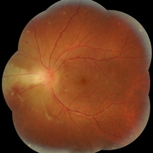
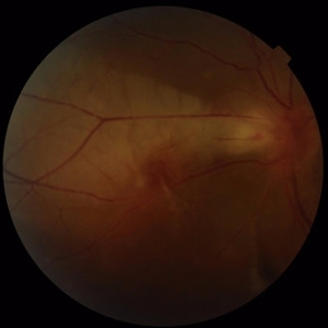
 Myelinated Nerve Fibre (MNF)
Jun 17 2023 by Harsh Vardhan Singh, MS
Fundus photograph of 32-year-old male having good best corrected visual acuity in both eyes with right eye having high myopia & MNF as incidental finding
Photographer: Dr Harsh Vardhan Singh, Assistant Professor, AIIMS, Guwahati
Condition/keywords: medullated nerve fibers, MNF, myelinated nerve fiber layer, myelinated nerve fibers, Nerve fiber layer arrangements, NFL
-
 "Starry Sky" Fundus in Vogt-Koyanaki-Harada Syndrome
Jan 10 2018 by Peter H. Tang, MD, PhD
Fluorescein angiography imaging of a 27-year-old male with acute inflammation as part of Vogt-Koyanagi-Harada Syndrome.
Imaging device: Optos California
Condition/keywords: chorioretinal inflammations, retina, uveitis, Vogt-Koyanagi-Harada
-
 'Wet Snow on Grapevines'
Apr 8 2019 by Gary R. Cook, MD, FACS
Fundus photograph of inflammatory deposits on vitreous fibrils, known as "Wet snow on grapevines" in a case of recurrent ocular toxoplasmosis.
Imaging device: Topcon VT-50
Condition/keywords: ocular toxoplasmosis, vitritis, wet snow on grapevines
-
 Acute Exudative Polymorphous Vitelliform Maculopathy Angio OD
Aug 27 2014 by Flavio A. Rezende, MD, PhD
45-year-old man with mild decrease in vision after strong headache. Fundus showing multiple deep irregular vitelliform lesions spread throughout entire posterior pole OU, forming a typical level of subretinal confluent lesions at the inferior retinal vascular arcades. No primary tumor or metastasis found.
Photographer: Eduardo Martins, Pontifícia Universidade Católica - Rio de Janeiro, Brazil
Imaging device: Topcon TRC 50EX
Condition/keywords: polymorphous exudative vitelliform maculopathy
-
 Acute Exudative Polymorphous Vitelliform Maculopathy Angio OS
Aug 27 2014 by Flavio A. Rezende, MD, PhD
45-year-old man with mild decrease in vision after strong headache. Fundus showing multiple deep irregular vitelliform lesions spread throughout entire posterior pole OU, forming a typical level of subretinal confluent lesions at the inferior retinal vascular arcades. No primary tumor or metastasis found.
Photographer: Eduardo Martins, Pontifícia Universidade Católica - Rio de Janeiro, Brazil
Imaging device: Topcon TRC 50EX
Condition/keywords: polymorphous exudative vitelliform maculopathy
-
 Acute Exudative Polymorphous Vitelliform Maculopathy Color OD
Aug 27 2014 by Flavio A. Rezende, MD, PhD
45-year-old man with mild decrease in vision after strong headache. Fundus showing multiple deep irregular vitelliform lesions spread throughout entire posterior pole OU, forming a typical level of subretinal confluent lesions at the inferior retinal vascular arcades. No primary tumor or metastasis found.
Photographer: Eduardo Martins, Pontifícia Universidade Católica - Rio de Janeiro, Brazil
Imaging device: Topcon TRC 50EX
Condition/keywords: polymorphous exudative vitelliform maculopathy
-
 Acute Exudative Polymorphous Vitelliform Maculopathy Color OS
Aug 27 2014 by Flavio A. Rezende, MD, PhD
45-year-old man with mild decrease in vision after strong headache. Fundus showing multiple deep irregular vitelliform lesions spread throughout entire posterior pole OU, forming a typical level of subretinal confluent lesions at the inferior retinal vascular arcades. No primary tumor or metastasis found.
Photographer: Eduardo Martins, Pontifícia Universidade Católica - Rio de Janeiro, Brazil
Imaging device: Topcon TRC 50EX
Condition/keywords: polymorphous exudative vitelliform maculopathy
-
 Acute Exudative Polymorphous Vitelliform Maculopathy Red Free OD
Aug 27 2014 by Flavio A. Rezende, MD, PhD
45-year-old man with mild decrease in vision after strong headache. Fundus showing multiple deep irregular vitelliform lesions spread throughout entire posterior pole OU, forming a typical level of subretinal confluent lesions at the inferior retinal vascular arcades. No primary tumor or metastasis found.
Photographer: Eduardo Martins, Pontifícia Universidade Católica - Rio de Janeiro, Brazil
Imaging device: Topcon TRC 50EX
Condition/keywords: polymorphous exudative vitelliform maculopathy
-
 Acute Exudative Polymorphous Vitelliform Maculopathy Red Free OS
Aug 27 2014 by Flavio A. Rezende, MD, PhD
45-year-old man with mild decrease in vision after strong headache. Fundus showing multiple deep irregular vitelliform lesions spread throughout entire posterior pole OU, forming a typical level of subretinal confluent lesions at the inferior retinal vascular arcades. No primary tumor or metastasis found.
Photographer: Eduardo Martins, Pontifícia Universidade Católica - Rio de Janeiro, Brazil
Imaging device: Topcon TRC 50EX
Condition/keywords: polymorphous exudative vitelliform maculopathy
-
---thumb.JPG/image-square;max$300,300.ImageHandler) Acute myeloid leukemia
Acute myeloid leukemia
Dec 9 2012 by Mallika Goyal, MD
Right eye of a 21-year-old gentleman with acute myeloid leukemia who is undergoing chemotherapy and has low platelet counts (17,000) shows multiple pre-retinal haemorrhages. Other eye has similar picture. There is no vascular occlusion or inflammation. Visual prognosis remains good with spontaneous resolution expected over few weeks.
Photographer: Mallika Goyal, MD, Apollo Health City, Hyderabad, India
Condition/keywords: preretinal hemorrhage
-
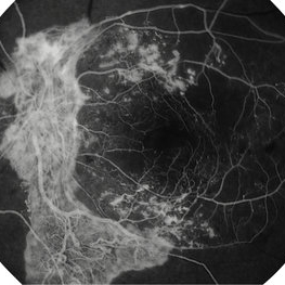
 Acute Posterior Multifocal Placoid Pigment Epitheliopathy
Jan 4 2019 by Cláudia Farinha
Composite image of both eyes of a 27-year-old male with APMPPE. In the fundus photograph, multiple yellowish placoid lesions are observed in the posterior pole in both eyes. The ICGA revealed more lesions than those observed in fundoscopy, and these were hypofluorescent through the angiogram as expected. The en face OCTA segmented at the level of the choriocapillaris revealed areas of ischemia in close correspondence with the hypofluorescent lesions (image superimposed in ICGA ). The OCT b-scan with superimposed flow shows disruption and hyperreflectivity of the external retinal layers in the affected areas and again the absence of flow in the choriocapillaris underneath. A systemic study was carried out to exclude other inflammatory and infectious causes of placoid retinochoroidopathy. The clinical picture resolved after approximately one month from the onset, without recurrence.
Photographer: Pedro Melo, Ophthalmology Department, Centro Hospitalar e Universitário de Coimbra, Coimbra Portugal
Condition/keywords: acute posterior multifocal placoid pigment epitheliopathy (APMPPE), white dot syndrome
-
 Acute Toxoplasmosis Neuroretinitis
Mar 15 2017 by Hamid Ahmadieh, MD
Color fundus photograph of the left eye of a 26-year-old man with clinical picture of acute neuroretinitis and serologic evidence of Toxoplasma gondii infection. Disc swelling, necrotizing retinitis with overlying vitreous inflammation, retinal vasculitis and localized exudative retinal detachment are visible.
Photographer: Solmaz Shahmohammad, Negah Eye Center, Tehran,Iran
Condition/keywords: color fundus photograph, exudative retinal detachment, neuroretinitis, retinal vasculitis, toxoplasmosis
-
 Acute Toxoplasmosis Neuroretinitis
Mar 15 2017 by Hamid Ahmadieh, MD
Color fundus photograph of the left eye of a 26-year-old man with clinical picture of acute neuroretinitis and serologic evidence of Toxoplasma gondii infection. Disc swelling, necrotizing retinitis with overlying vitreous inflammation, retinal vasculitis and localized exudative retinal detachment are visible.
Photographer: Solmaz Shahmohammad, Negah Eye Center, Tehran,Iran
Condition/keywords: color fundus photograph, exudative retinal detachment, neuroretinitis, retinal vasculitis
-
 Advanced Proliferative Diabetic Retinopathy With Fibrovascular Proliferation
Jan 4 2019 by Isha Agarwalla
A 29-year-old female with a long-standing history of diabetes mellitus presented with a fibrovascular membrane (FVM) at the viteroretinal interface due to underlying inflammation and angiogenesis induced by ischemia. FVM involved the disc and extended towards the superior and inferior arcades along with extensive capillary drop out areas due to micro aneurysms.
Condition/keywords: fibrovascular proliferation, proliferative diabetic retinopathy (PDR)
-
 Advanced Proliferative Diabetic Retinopathy With Fibrovascular Proliferation
Jan 4 2019 by Isha Agarwalla
A 29-year-old female with a long-standing history of diabetes mellitus presented with a fibrovascular membrane(FVM) at the viteroretinal interface due to underlying inflammation and angiogenesis induced by ischemia. FVM involved the disc and extended towards the superior and inferior arcades along with extensive capillary drop out areas due to micro aneurysms.
Condition/keywords: fibrovascular proliferation, fluorescein angiogram (FA), proliferative diabetic retinopathy (PDR)
-
 AMPPPE
Apr 17 2013 by Howard Schatz, MD
AMPPPE, inflamed.
Condition/keywords: acute posterior multifocal placoid pigment epitheliopathy (APMPPE)
-
 Anemic Retinopathy Related Retinal Hemorrhages
Nov 5 2019 by Chinmayi Vyas
Anemic retinopathy related retinal hemorrhages in a 24 years old male with Hb of 4.2gm/ dl. The manifestations of anemic retinopathy are nonspecific and may closely simulate hypertensive or diabetic retina. Retinal changes in anemia are cotton wool spots, venous tortuosity, and hemorrhages which may be present at all levels of the retina and choroid. All retinal hemorrhages can occur when Hb falls below 8 g/100 ml or if the platelet count falls below 50,000/cumm. The combination of severe anemia and thrombocytopenia is likely to produce retinal hemorrhages. The Roth’s spots or white centre hemorrhages are typically associated with bacterial endocarditis , anemia and other systemic conditions. The white center is suspected to represents focal ischemia, inflammatory or infectious infiltrate, fibrin or accumulation of neoplasticism cells.
Photographer: Dr Chinmayi Vyas
Condition/keywords: retinal hemorrhage
-
 Anemic Retinopathy Related Retinal Hemorrhages
Nov 5 2019 by Chinmayi Vyas
Anemic retinopathy related retinal hemorrhages in a 24 years old male with Hb of 4.2gm/ dl. The manifestations of anemic retinopathy are nonspecific and may closely simulate hypertensive or diabetic retina. Retinal changes in anemia are cotton wool spots, venous tortuosity, and hemorrhages which may be present at all levels of the retina and choroid. All retinal hemorrhages can occur when Hb falls below 8 g/100 ml or if the platelet count falls below 50,000/cumm. The combination of severe anemia and thrombocytopenia is likely to produce retinal hemorrhages. The Roth’s spots or white centre hemorrhages are typically associated with bacterial endocarditis , anemia and other systemic conditions. The white center is suspected to represents focal ischemia, inflammatory or infectious infiltrate, fibrin or accumulation of neoplasticism cells.
Photographer: Dr Chinmayi Vyas, Nethradhama superspeciality eye hospital , banglore, india
Imaging device: Eidon fundus imaging
Condition/keywords: anaemic retinopathy
-
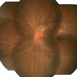
---thumb.jpg/image-square;max$300,300.ImageHandler) ARMD RPE Defect / Myelinated NFL
ARMD RPE Defect / Myelinated NFL
Jan 9 2014 by David Callanan, MD
ARMD RPE defect , myelinated nerve fiber layer in a 47-year-old male patient.
Condition/keywords: myelinated nerve fiber layer, retinal pigment epithelium (RPE) defect
-
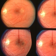
 ARN (#1) Initial Photo
May 27 2019 by John S. King, MD
60-year-old African American female who had been treated for iridocyclitis for at least a week sent in for vitritis and a nasal fundus lesion. Complaints included redness, floaters, photophobia, and decreased vision. Husband had recent shingles. Acuity was 20/60-2 with IOP of 12, and small KP in Art's triangel, 1-2+ a/c cell, 2-3+ ant vit cell, diffuse arteriolar sheathing, multiple areas of retinal whitening in periphery and mid-periphery (see Photo #1). PCR of a/c was performed, and intravitreal GCV administered, and VACV 2g qid and ASA started.... PCR positive for HZV, pred taper was started two days after presentation as the infection had begun to stablize..... Five days from presentation the vision was 20/60, inflammation and areas of retinal whitening had improved (see Photo #2).... One week later acuity was 20/30, the a/c was quiet and KP resolved; ant vitreous cell decreased; and there was further improvement in retinal appearance without any signs of retinal holes or detachment; she is now on low dose maint VACV (see photo#3)
Photographer: Maysee Yang
Imaging device: Optos CA
Condition/keywords: acute retinal necrosis, Herpes zoster
 Loading…