Search results (289 results)
-
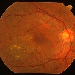
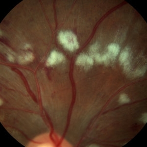
 Diabetes NPDR
Mar 29 2013 by Henry J. Kaplan, MD
NPDR.
Condition/keywords: cotton wool spots, nonproliferative diabetic retinopathy
-
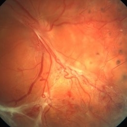 Wolf Jaw Detachment
Mar 11 2013 by Jones Jackie
30-year-old male with 19 year history of Diabetes Mellitus. Severe traction retinal detachment with proliferative diabetic retinopathy.
Photographer: Keith Anderson, Edina Retina Consultants, Edina Minnesota
Condition/keywords: diabetic mellitus
-
 Diabetic Retinal Hemorrhages in Proliferative Diabetes
Sep 10 2012 by James B. Soque, CRA, OCT-C, COA, FOPS
Fundus Photo of Severe Proliferative Diabetic with Retinal Hemorrhages, Left eye, scattered laser treatment. View: 50 Degrees
Photographer: James Soque, CRA, COA, Island Retina, Shirley, NY
Imaging device: Topcon TRC 50 DX
Condition/keywords: proliferative diabetic retinopathy (PDR)
-
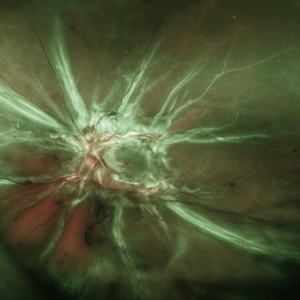
 Oliferative Diabetic Retinopathy, Neovascularization of Disc
Mar 28 2018 by awaneesh m upadhyay, MBBS, DNB
Fundus photograph of right eye of a 56-year-old gentleman having diabetes, hypertension and chronic kidney disease.
Photographer: Dr Awaneesh Upadhyay
Imaging device: Zeiss
Condition/keywords: proliferative diabetic retinopathy (PDR)
-
---thumb.jpg/image-square;max$300,300.ImageHandler) Diabetes with Cortical Cataract
Diabetes with Cortical Cataract
Apr 4 2014 by H. Michael Lambert, MD
Diabetes. Cortical Spoking
Photographer: Donald Lowd
Condition/keywords: cataract, diabetes
-
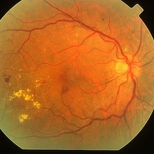
 Diabetic Retinopathy, CSME, Color Fundus Photo
Mar 18 2015 by James B. Soque, CRA, OCT-C, COA, FOPS
A 58-year-old diabetic male with a longstanding history of diabetic eye disease. Left eye color fundus photo shows extensive CSME, Clinically Significant Macular Edema, with deposits of hard exudates at fixation. There is extensive scattering of hard exudates and sheathing of the vessels.
Photographer: James B Soque, CRA COA
Imaging device: Topcon TRC 50 DX, OIS 5 MP Camera, MERGE software
Condition/keywords: background diabetic retinopathy (BDR), creamy yellow exudates, diabetes, exudates over the posterior pole, neovascularization of the disc (NVD), vessel sheathing
-
 Diabetes HRC-PDR
Mar 29 2013 by Henry J. Kaplan, MD
NVD on the optic disc extended superiorly and temporally.
Condition/keywords: neovascularization of the disc (NVD)
-
 FFA - PDR
Mar 30 2018 by Lanin Chen
Fundus fluorescein angiography photo of the left eye of a 62-year-old woman with history of Type 2 diabetes mellitus since 20 years showing proliferative diabetic retinopathy.
Photographer: Lanin Chen
Condition/keywords: fundus autofluorescence (FAF), proliferative diabetic retinopathy (PDR)
-
 MPC for CSME
Mar 29 2013 by Henry J. Kaplan, MD
Right after MPC for CSME in diabetes (before the introduction of anti-VEGFs).
Condition/keywords: clinically significant macular edema (CSME), diabetic macular edema, multifocal chorioretinitis (MCP)
-
 Melanocytoma Case 2
Oct 5 2012 by Ronald C. Gentile, MD
Melanocytoma of the optic disc in a patient with diabetes. The elevated, black melanocytoma is seen impinging on the optic nerve with some unrelated nonproliferative diabetic retinopathy. Patient had a corresponding inferior visual field defect.
Photographer: The New York Eye & Ear Infirmary Department of Medical Imaging
Condition/keywords: melanocytoma, nonproliferative diabetic retinopathy
-
 Advanced PDR
Sep 29 2012 by Hamid Ahmadieh, MD
Color fundus photograph of a 32-year-old man with insulin-dependent diabetes mellitus and regressed advanced PDR with severe fibrous proliferation and traction retinal detachment sparing the macula.
Photographer: Hamid Ahmadieh, MD; Ophthalmic Research Center, Labbafinejad Medical Center, Shahid Beheshti University of Medical Sciences
Condition/keywords: fibrous proliferation, tractional retinal detachment
-
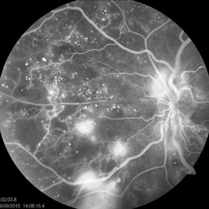
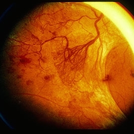
 Proliferative Diabetic Retinopathy
Oct 15 2012 by Susanna S. Park, MD, PhD
Fluorescein angiogram of the left eye of a 65 year old woman with diabetes mellitus showing nasal peripheral retinal capillary dropout and neovascularization of the disc. Scattered retinal microaneurysms are also noted
Photographer: Ellen Redenbo, University of California Davis Eye Center
Imaging device: Optos
Condition/keywords: proliferative diabetic retinopathy (PDR)
-
 Diabetic Retinopathy, CSME, Exudates, NVD, Color Fundus Photo, Montage
Mar 18 2015 by James B. Soque, CRA, OCT-C, COA, FOPS
A 58-year-old diabetic male with a longstanding history of diabetic eye disease. Left eye color fundus photo shows extensive CSME, Clinically Significant Macular Edema, with deposits of hard exudates at fixation. There is extensive scattering of hard exudates and sheathing of the vessels.
Photographer: James B Soque, CRA COA
Imaging device: Topcon TRC 50 DX, OIS 5 MP Camera, MERGE software
Condition/keywords: background diabetic retinopathy (BDR), creamy yellow exudates, diabetes, exudates over the posterior pole, neovascularization of the disc (NVD), vessel sheathing
-
 Advanced PDR
Sep 29 2012 by Hamid Ahmadieh, MD
Color fundus photograph of a 32-year-old man with insulin-dependent diabetes mellitus and regressed advanced PDR with severe fibrous proliferation and traction retinal detachment sparing the macula.
Photographer: Hamid Ahmadieh, MD; Ophthalmic Research Center, Labbafinejad Medical Center, Shahid Beheshti University of Medical Sciences
Condition/keywords: fibrous proliferation, tractional retinal detachment
-
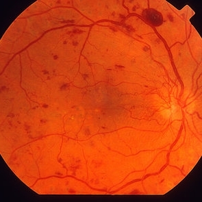
 Proliferative Diabetic Retinopathy
Jul 9 2012 by George W. Aylward, MD, FRCS, FRCOphth
Severe proliferative diabetic retinopathy in a patient with long standing insulin dependent diabetes and no ophthalmic treatment.
Condition/keywords: diabetic mellitus, insulin dependent
-
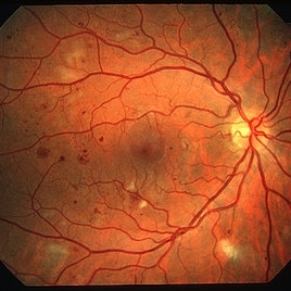
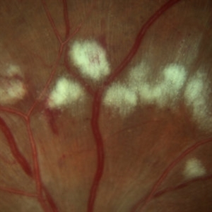
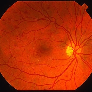 Diabetes NPDR
Mar 29 2013 by Henry J. Kaplan, MD
Moderate NPDR.
Condition/keywords: nonproliferative diabetic retinopathy
-
 Vitreous Hemorrhage
Jul 10 2018 by Karen Panzegrau
SD-OCT of a 35-year-old female presenting with a vitreous hemorrhage of her left eye. Patient has active proliferative diabetic retinopathy, as well as a completed posterior vitreous detachment in the left eye.
Photographer: Karen Panzegrau
Condition/keywords: diabetes, Heidelburg Spectralis, left eye, optical coherence tomography (OCT), posterior vitreous detachment, proliferative diabetic retinopathy (PDR), vitreous hemorrhage
-
 Diabetes NPDR
Mar 29 2013 by Henry J. Kaplan, MD
Moderated NPDR.
Condition/keywords: diabetic mellitus, nonproliferative diabetic retinopathy
-
 Advanced Active PDR
Mar 29 2013 by Henry J. Kaplan, MD
Large active NVEs with fibrous proliferations in diabetes.
Condition/keywords: fibrous proliferation, neovascularization (NV)
-
 Diabetic Retinopathy
Oct 2 2013 by Jerald A. Bovino, MD
This patient with long standing diabetes has peripheral non-perfusion.
Condition/keywords: retinal ischemia
-
 Cotton Wool Spots
Mar 1 2014 by Homayoun Tabandeh, MD, FASRS
Cotton wool spots in a patient with hypertension and diabetes
Condition/keywords: cotton wool spots, diabetic retinopathy, hypertensive retinopathy
-
 Diabetic Retinopathy Optic Nerve Edema, Fluorescein Angiogram, Stereo
Apr 11 2015 by James B. Soque, CRA, OCT-C, COA, FOPS
Optic Nerve Edema and Leakage on fluorescein angiography in this 48-year-old patient with a 10 year history of diabetes. 50 degree stereo photo fluorescein angiogram.
Photographer: James B. Soque, CRA, COA
Imaging device: Topcon TRC 50 DX, OIS 5 MP Digital Camera, MERGE Software
Condition/keywords: background diabetic retinopathy (BDR), diabetes, disc leakage, fluorescein leakage, optic disc swelling, optic nerve edema, stereo pair
-
 Diabetic Tractional Retinal Detachment
May 19 2014 by John W. Kitchens, MD
Severe PDR with non perfusion and a tractional retinal detachment.
Photographer: Ed Slade
Imaging device: Optos 200Tx
Condition/keywords: diabetes
-
 Cotton Wool Spots
Mar 1 2014 by Homayoun Tabandeh, MD, FASRS
Cotton wool spots in a patient with hypertension and diabetes.
Condition/keywords: cotton wool spots, diabetic retinopathy, hypertensive retinopathy
 Loading…