Search results (289 results)
-
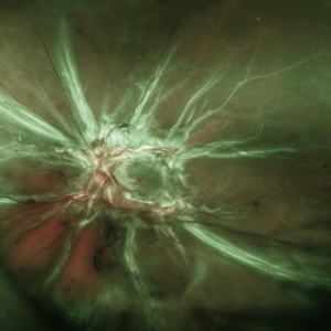
 Tractional Retinal Detachment
Dec 4 2019 by Janet Brazil
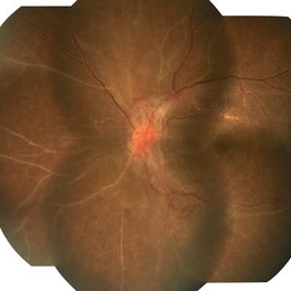
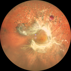
Fundus photograph of a 32-year-old female with severe end-stage diabetic tractional retinal detachment.
Photographer: Janet Atkinson, Eye Associates of New Mexico, Albuquerque, NM
Imaging device: Topcon TRC- 50EX
Condition/keywords: diabetes, proliferative diabetic retinopathy (PDR), tractional retinal detachment
-
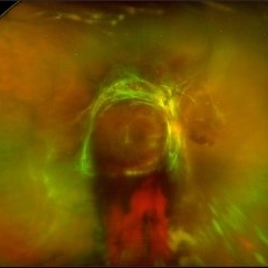
 Candy Stripe Sign
Mar 30 2023 by pedro fernandes souza neto
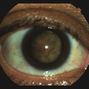
Candy Stripe Sign, patient with proliferative diabetic retinopathy progressing to vitreous hemorrhage and subsequently to ghost cell glaucoma.
Photographer: Marlos Henrique Oliveira Junior, Federal University of Bahia.
Condition/keywords: dehemoglobinized hemorrhage, diabetes, diabetic glaucoma
-
 Diabetic traction retinal detachment
Jan 9 2023 by JORGE SOBERANES
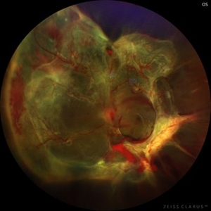
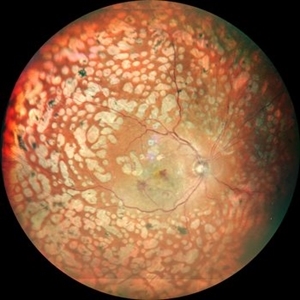
Proliferative diabetic retinopathy with extensive traction retinal detachment in a patient with type 1 diabetes mellitus.
Photographer: Dr. Jorge I. Soberanes, Asociación para Evitar la Ceguera en México.
Imaging device: Zeiss Clarus 700
Condition/keywords: Retinal Detachment, tractional retinal detachment
-
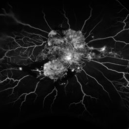
 Proliferative Diabetic Retinopathy
Jul 15 2022 by Gabriel Costa Andrade, PhD
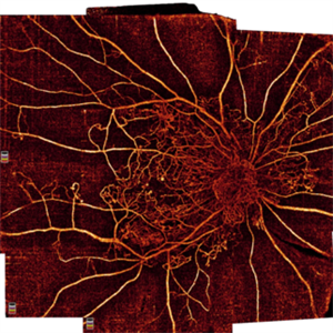
Fundus angiography of an 22-year-old man with proliferative diabetic retinopathy and macular ischemia.
Photographer: Dr Gabriel Andrade
Condition/keywords: Diabetes
-
 Proliferative Diabetic Retinopathy
Oct 16 2021 by Timur Shaimov
32 y.o. female with Type 1 Diabetes with no glucose compensation for several years. A manual montage of several 8x8 mm OCT angiograms were obtained for this Widefield OCTA image.
Photographer: Timur Shaimov
Imaging device: RTVue xR Avanti
Condition/keywords: OCT Angiography, proliferative diabetic retinopathy (PDR)
-
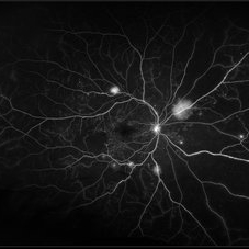
 Proliferative Diabetic Retinopathy with Severe Ischemia
Nov 30 2023 by Gabriel Costa Andrade, PhD
Ultra-widefield fluorescein angiography of the right eye of a 47 year old woman with diabetes mellitus showing macular and nasal retinal capillary dropout and neovascularization of the disc and temporal vascular arcades.
Photographer: Gabriel Andrade
Imaging device: Optos California
Condition/keywords: Diabetic Retinopathy
-
 Venous Beading
Apr 30 2021 by Shivani Reddy, MD
This is a fluorescein angiogram image capturing a beautiful example of different stages of venous beading in diabetic retinopathy all in one frame. This patient also has various microangiopathic findings including microaneurysms, venous loops and capillary dropout. This patient is a 41 y/o male with a history of type 1 diabetes, presenting for his first eye exam in years.
Imaging device: Optos FA
Condition/keywords: capillary dropouts, nonproliferative diabetic retinopathy, proliferative diabetic retinopathy (PDR), retinal ischemia, venous beading
-
 Advanced Proliferative Diabetic Retinopathy
Nov 4 2017 by Hamid Ahmadieh, MD
Merged color fundus photograph of the left eye of a 30-year-old woman with type1 diabetes since childhood. Note laser scars, severe fibrous proliferation, traction RD and macular dragging.
Photographer: Shabnam Poureh, Negah Eye Center, Tehran, Iran
Condition/keywords: color fundus photograph, diabetes, fibrous proliferation, proliferative diabetic retinopathy (PDR), severe traction
-
 Advanced Proliferative Diabetic Retinopathy With Fibrovascular Proliferation
Jan 4 2019 by Isha Agarwalla
A 29-year-old female with a long-standing history of diabetes mellitus presented with a fibrovascular membrane (FVM) at the viteroretinal interface due to underlying inflammation and angiogenesis induced by ischemia. FVM involved the disc and extended towards the superior and inferior arcades along with extensive capillary drop out areas due to micro aneurysms.
Condition/keywords: fibrovascular proliferation, proliferative diabetic retinopathy (PDR)
-
 Annular Tractional Retinal Detachment
Jul 4 2024 by Hector Gabriel Moreno Solano, MD, MHA
52-year-old Hispanic female patient with a diagnosis of type II diabetes mellitus of 15 years of evolution, comes to the retina service for progressive visual loss in the right eye (single functional eye) with visual acuity of 20/100, Fundus examination reveals laser-modified proliferative diabetic retinopathy with activity + annular tractional retinal detachment with macular involvement.
Photographer: Hector Gabriel Moreno Solano, MD, MHA, HGZ #20 IMSS Puebla.
Imaging device: Mirante
Condition/keywords: macular detachment, proliferative diabetic retinopathy (PDR), tractional retinal detachment
-
 Central Retinal Artery Occlusion
Apr 20 2018 by Kim Barrett
64-year-old female woke with no vision in her right eye. This image was taken at 6:11 minutes and the vessels have not filled. Patient has been treated with PRP laser and anti-VEGF therapy. Current vision is CF @ 2 ft.
Photographer: Kim Barrett C.O.A.
Imaging device: Heidelberg
Condition/keywords: central retinal artery occlusion (CRAO), diabetes, hypertension, smoker, uncontrolled
-
 CNVM in Pan-retinal Photocoagulated Patient
Dec 30 2020 by ASRS Staff
Wide fundus photograph of 65-year-old, female, diabetic patient.
Imaging device: Nidek Mirante
Condition/keywords: age-related macular degeneration (AMD), diabetes, pan-retinal photocoagulation (PRP)
-
 Detached NVE During PVD induction
Apr 27 2018 by Michael J. Koss, MD, PhD, MBA
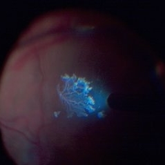
A 73-year-old woman with macular pucker underwent a pars plana vitrectomy with membrane peeling. Additionally the patient suffers from diabetic retinopathy after being diagnosed with type 2 diabetes mellitus sixteen years ago. Prior to the procedure she was treated with a series of intravitreal Bevacizumab-injections due to diabetic macular edema. There was no history of a proliferative DRP. During the vitrectomy a branch of an obliterated NVE spontaneously detached and floated freely in the vitreous. The 3D shot was captured via Alcon’s NGENUITY® 3D Visualization System in form of photograph and video providing an outstandingly detailed image of the branched NVE.
Photographer: Michael Koss, Augenzentrum Nymphenburger Hoefe
Imaging device: Alcon’s NGENUITY® 3D Visualization System
Condition/keywords: diabetes, diabetic retinopathy, neovascularization elsewhere (NVE), pars plana vitrectomy (PPV), PVD induction
-
 Diabetes HRC-PDR
Mar 29 2013 by Henry J. Kaplan, MD
NVD on the optic disc extended superiorly and temporally.
Condition/keywords: neovascularization of the disc (NVD)
-
 Diabetic Retinopathy, CSME, Exudates, NVD, Color Fundus Photo, Montage
Mar 18 2015 by James B. Soque, CRA, OCT-C, COA, FOPS
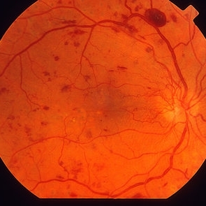
A 58-year-old diabetic male with a longstanding history of diabetic eye disease. Left eye color fundus photo shows extensive CSME, Clinically Significant Macular Edema, with deposits of hard exudates at fixation. There is extensive scattering of hard exudates and sheathing of the vessels.
Photographer: James B Soque, CRA COA
Imaging device: Topcon TRC 50 DX, OIS 5 MP Camera, MERGE software
Condition/keywords: background diabetic retinopathy (BDR), creamy yellow exudates, diabetes, exudates over the posterior pole, neovascularization of the disc (NVD), vessel sheathing
-
 Diabetic Tractional Retinal Detachment
May 19 2014 by John W. Kitchens, MD
Severe PDR with non perfusion and a tractional retinal detachment.
Photographer: Ed Slade
Imaging device: Optos 200Tx
Condition/keywords: diabetes
-
 Diabetic Tractional Retinal Detachment
Jan 23 2019 by Olivia Rainey
Ultra-wide field pseudocolor image of an 43-year-old female with a diabetic tractional retinal detachment and a vitreous hemorrhage affecting her right eye.
Photographer: Olivia Rainey
Imaging device: Optos
Condition/keywords: diabetes, diabetic traction detachment, Optos, pan-retinal photocoagulation (PRP), proliferative diabetic retinopathy (PDR), pseudocolor, ultra-wide field imaging, vitreous hemorrhage
-
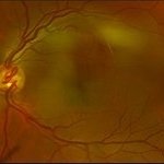 HTN Retinopathy with Pre-Papillary Vascular Loop OS
Jun 4 2018 by Hosam Attia, MD
Close-up color fundus photograph of 53-year-old, African American male with history of diabetes, hypertension, depicting chronic hypertensive retinopathy changes and unilateral pre-papillary vascular loop OS.
Imaging device: Optos California
Condition/keywords: congenital prepapillary vascular anomaly, congenital prepapillary vascular loop, prepapillary vascular loop
-
 Maternally-Inherited Diabetes and Deafness (MIDD) Syndrome
Jan 12 2025 by Niloofar Piri, MD
Fundus Autofluorescence image of right posterior pole in a 43 year old female who was referred for diabetic retinopathy evaluation, demonstrated multiple patches of hypoautrofluorescence surrounding the nerve and fovea. Please note that central fovea is spared. Granular hyper and hypoauto fluorescence is present in the macula and peripapillary region. She was noted to have hearing loss as well and after further evaluation was diagnosed with MIDD syndrome.
Condition/keywords: Maternally inherited diabetes and deafness (MIDD), Maternally-inherited-diabetes-and-deafness-(MIDD) syndrome, Mitochondrial Disorder
-
 MIDD (Maternally Inherited Diabetes and Deafness) - Left AF
Nov 30 2024 by John S. King, MD
Both right and left eyes have symmetrical ring of mottled hypo/hyper AF around the fovea and disc. The HyperAF areas correspond to RPE deposits on OCT as well as areas of blockage on FA, and drusenoid deposits seen on fundus photos 57 yo WF referred for AMD vs Pattern Dystrophy that was diagnosed 10 years ago. Reported some slow progressive vision loss in both eyes for distance and near. Denies nyctalopia or hemeralopia. Background medical history includes HTN, CVD, and DM. No family history of eye problems. Denied pentosan use. Anterior segment showed moderate cataracts (OD>OS). Posterior segment exam showed macular changes and mild NPDR. The macular appearance showed a symmetrical, paramacular ring of fleck-like drusenoid material with some faint focal areas of RPE hyperplasia. Fundus Photos, AF, OCT were performed as well as a gene test. Further questioning showed revealed that her mother and maternal grandmother had both diabetes mellitus and sensorineural hearing loss. The patient developed diabetes in her teens, and some high frequency hearing loss in her early twenties. She had not had a previous genetic test or diagnosis of MIDD. Gene testing is pending for the mitochondrial component. Invitae's retinal panel, which does not include mitochondrial disorders, only showed a variant of uncertain significance, HMCN1. I discussed this case with Dr. Freund, and it is similar to a the case report : Inoue M, Kiss S, Freund KB. MACULAR PIGMENT RINGS AS THE PRESENTING FINDING OF MITOCHONDRIAL MYOPATHY, ENCEPHALOPATHY, LACTIC ACIDOSIS, AND STROKELIKE EPISODES. Retin Cases Brief Rep. 2015 Fall;9(4):260-4. doi: 10.1097/ICB.0000000000000182. PMID: 26200388.
Photographer: Grace Melton and Carley Gunn
Imaging device: Clarus
Condition/keywords: Macular Dystrophy, Maternally Inherited Diabetes and Deafness, MIDD, Mitochondrial Disorder
-
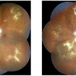 NPDR With Myelinated Nerve Fibers
Nov 5 2018 by Diva Kant Misra, MBBS, DO, DNB, MNAMS, FVRS
Bilateral montage funds photo images of a 56-year-old diabetic patient showing signs of NPDR along with myelinated nerve fibers.
Photographer: Hiteshwar Saikia
Condition/keywords: diabetes, hard exudates, myelinated nerve fibers, nonproliferative diabetic retinopathy
-
 Proliferative Diabetic Retinopathy
Mar 1 2025 by Neeket R. Patel, MD
A fundus photograph of a 28-year-old male monocular patient diagnosed with proliferative diabetic retinopathy, who is highly motivated to pursue retinal surgery. This case presents a unique challenge in both management and communication, given the patient's strong desire for intervention and the guarded prognosis for visual improvement.
Condition/keywords: Diabetes, membranes, retinopathy, Traction retinal detachment
-
 Proliferative Diabetic Retinopathy
Jan 29 2021 by Olivia Rainey
Ultra-widefield fluorescein angiogram of a 65-year-old male with proliferative diabetic retinopathy affecting his right eye. The patient's diabetic retinopathy has progressed significantly since he was last seen in 2014. It was recommended to begin antiVEGF to control DME followed by laser treatment OU.
Photographer: Olivia Rainey, OCT-C, COA
Imaging device: Optos California
Condition/keywords: anti-VEGF, diabetes, diabetic macular edema, fluorescein angiogram (FA), fluorescein leakage, neovascularization (NV), neovascularization elsewhere (NVE), non-perfusion, Optos, proliferative diabetic retinopathy (PDR), ultra-wide field imaging
-
 Proliferative Diabetic Retinopathy
Aug 16 2022 by Donnie Willis
51 yo female. Uncontrolled Diabetes. Active PDR.
Photographer: Donnie Willis, Tennessee Retina
Imaging device: Optos
Condition/keywords: capillary dropouts, Diabetes, FA, fluorescein angiogram (FA), Optos, proliferative diabetic retinopathy (PDR), vitreomacular traction (VMT)
-
 Proliferative Diabetic Retinopathy
Jul 9 2012 by George W. Aylward, MD, FRCS, FRCOphth
Severe proliferative diabetic retinopathy in a patient with long standing insulin dependent diabetes and no ophthalmic treatment.
Condition/keywords: diabetic mellitus, insulin dependent
 Loading…