Search results (34 results)
-
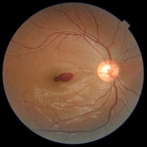
 Valsalva Retinopathy
Nov 18 2022 by Niloofar Piri, MD
Sudden vision loss immediately after severe vomiting. Color fundus photo demonstrates large sub ILM hemorrhage consistent with valsalva retinopathy.
Photographer: Sean Kelso, Saint Louis University
Condition/keywords: SUB ILM hemorrhage, sub internal limiting membrane haemorrhage, valsalva retinopathy
-
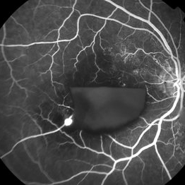
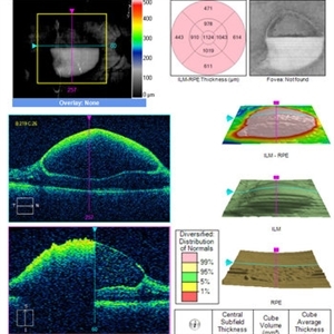
 Horizontal OCT Scan of Sub ILM Hemorrhage
Mar 8 2017 by Manish Nagpal, MD, FRCS (UK), FASRS
Patient with a macroaneurysm leading to a sub ILM hemorrhage near fovea showing an interesting horizontal scan passing through the central area.
Photographer: pranita chaudhary
Condition/keywords: hemorrhage, macroaneurysm
-
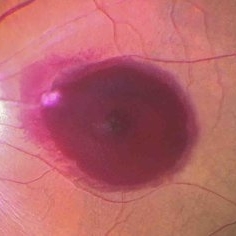
 Macroaneurysm With Sub ILM Hemorrhage
Mar 8 2017 by Manish Nagpal, MD, FRCS (UK), FASRS
Patient with a macroaneurysm leading to a sub ILM hemorrhage near fovea.
Photographer: Pranita Chaudhary
Condition/keywords: hemorrhage, macroaneurysm
-
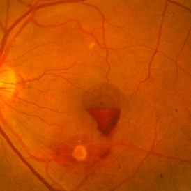
 RAMA with Sub ILM Hemorrhage
Jan 31 2018 by John S. King, MD
73-year-old with well controlled diabetes and hypertension presented with a month onset of acute central scotoma; CF 5'
Photographer: Stacey
Imaging device: Cirrus
Condition/keywords: ruptured macroaneurysm, sub-inner limiting membrane hemorrhage
-
 Sub ILM Hemorrhage
Jan 12 2022 by Manish Nagpal, MD, FRCS (UK), FASRS
Intraoperative view of a non clearing sub ILM hemorrhage over the macula with partly de-hemoglobinized blood.
Photographer: Manish Nagpal, Retinal Foundation, Ahmedabad, India
Imaging device: Sony PMW -10 MD surgical camera
Condition/keywords: sub internal limiting membrane haemorrhage, subILM hemorrhage
-
 Sub ILM Hemorrhage
May 19 2023 by Rahul Bhatia, MS, DNB
A 10-year-old male with Aplastic Anemia presented to Retina Clinic. Fundus Photograph and OCT line scan suggestive of Sub ILM Hemorrhage
Photographer: Dr Rahul Bhatia, LHMC, Delhi, India
Imaging device: Iphone
Condition/keywords: sub internal limiting membrane haemorrhage
-
 Valsalva Retinopathy
Nov 18 2022 by Niloofar Piri, MD
21 yo female presented with decaresed central vision and scotoma immediately after severe vomiting. Color fundus phtograph demonstrates large sub ILM layered hemorrhage in the macula consistent with valsalva retinopathy. Notice the sacttered blot retinal hemorrhages in mid-periphery.
Photographer: Rocio Bentivegna, MD, Saint Louis University
Condition/keywords: sub ILM hemorrhage, valsalva retinopathy
-
 Small subILM Hemorrhage
Oct 26 2019 by Navneet Mehrotra, DNB
44-year-old hypertensive male with sudden decrease in vision showing small sub ILM hemorrhage at macula.
Photographer: Navneet Mehrotra
Imaging device: NidekRS330
Condition/keywords: hypertension, subILM hemorrhage
-
 Fishing Fundus
Jul 16 2025 by Moazzam Parvez
Fundus photograph of a 31 year old woman with sub ILM hemorrhage following her yoga sessions which involves breath holding exercises .
Photographer: Moazzam Parvez , Netralayam , Kolkata
Imaging device: Topcon Maestro 2
Condition/keywords: SUB ILM hemorrhage, valsalva retinopathy
-
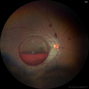
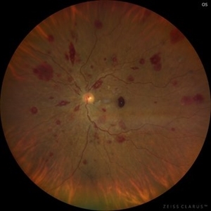 Leukemic Retinopathy
Nov 27 2024 by Ramses Rosales-Diaz
Fundus photograph of a 48-year-old woman with venous dilatation and tortuosity, flame-shaped and intraretinal hemorrhages, Roth spots and sub-ILM hemorrhage. Her complete blood count reports 425,540 lymphocytes/microliter, and the blood smear reveals Gumprecht shadows and numerous lymphocytes with nuclei exhibiting hypercondensed chromatin. She is diagnosed with chronic lymphocytic leukemia and receives appropriate treatment from the hematology team
Photographer: Ramses Rosales-Diaz, Asociación Para Evitar la Ceguera en México
Imaging device: Clarus 700
Condition/keywords: leukemia, sub ILM hemorrhage, white centered retinal hemorrhage (Roth Spot)
-
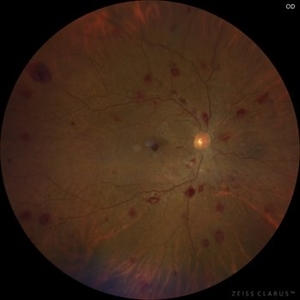 Leukemic Retinopathy
Nov 27 2024 by Ramses Rosales-Diaz
Fundus photograph of a 48-year-old woman showing venous dilatation and tortuosity, flame-shaped hemorrhages, intraretinal hemorrhages, sub-ILM hemorrhages, and Roth spots. Her complete blood count shows 425,540 lymphocytes/microliter, and the blood smear reveals Gumprecht shadows along with numerous lymphocytes with hypercondensed chromatin in their nuclei. She is diagnosed with chronic lymphocytic leukemia and receives appropriate treatment from the hematology team.
Photographer: Ramses Rosales-Diaz, Asociación Para Evitar la Ceguera en México
Imaging device: Zeiss Clarus 700
Condition/keywords: leukemia, sub ILM hemorrhage, white centered retinal hemorrhage (Roth Spot)
-
 RAMA with Sub ILM Hemorrhage
Jan 31 2018 by John S. King, MD
73 -year-old with well controlled diabetes and hypertension presented with a month onset of acute central scotoma; CF 5'; FA shows pooling in the aneurysm, blockage by dehemoglobinized heme, some diabetic changes and some IRMAs likely from old vein occlusion (s)
Photographer: Stacey
Imaging device: Cirrus
Condition/keywords: ruptured macroaneurysm, sub-inner limiting membrane hemorrhage
-
 RAMA with Sub ILM Hemorrhage
Jan 31 2018 by John S. King, MD
73-year-old with well controlled diabetes and hypertension presented with a month onset of acute central scotoma; CF 5'; SUB-ILM vs subyaloid elevation
Photographer: Stacey
Imaging device: Cirrus
Condition/keywords: ruptured macroaneurysm, sub-inner limiting membrane hemorrhage
-
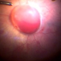
 Retinal Artery Macro-aneurysms
Jul 19 2024 by Anjana Mirajkar, MS Ophthalmology
An intra operative still of LE showing a retinal artery macro aneurysm causing a sub hylaoid and sub ILM hemorrhage.
Photographer: Dr. Anjana Mirajkar -Retina Foundation, Ahmedabad
Imaging device: Mirante-Nidek
Condition/keywords: retinal arterial macroaneurysm, sub hyaloid hemorrhage, sub internal limiting membrane haemorrhage
-
 Shaken Baby Syndrome
Feb 20 2022 by Imren Akkoyun, MD, FACS
Fundus photograph of an 6 months-old girl. Fundus exam revealed retinal hemorrhages involving multiple layers with subhyaloidal and sub ILM hemorrhage involving optic disc and macula.
Photographer: Imren Akkoyun, MD, FACS;Baskent University Faculty of Medicine Department of Ophthalmology
Imaging device: Zeiss, Operating Microscope
Condition/keywords: Shaken-Baby-Syndrome
-
 Sub ILM Dehaemoglobinised Hemorrhage With Retinal Detachment
Jan 16 2025 by Anand Temkar
A 39 year old male was referred to us with this presentation after a month of his first vitrectomy surgery done for VH e/w. His serum homocysteine was raised but MRI brain was within normal limits. We can see the sub ILM dehaemoglobinised hemorrhage (supero-temporal to macula) and Retinal detachment (inferiorly and nasally).
Photographer: Dr.Anand Temkar- Retina Foundation, Ahmedabad
Imaging device: Mirante
Condition/keywords: dehemoglobinized hemorrhage, Retinal Detachment, SUB ILM hemorrhage
-
 Sub ILM Dehaemoglobinised Hemorrhage With Retinal Detachment in Vitrectomised Eye
Jan 16 2025 by Anand Temkar
A 39 yrs old male was referred to us with this presentation after a month of his first vitrectomy surgery done for VH e/w. His serum homocysteine was raised but MRI brain was within normal limits. We can see the sub ILM dehaemoglobinised hemorrhage (supero-temporal to macula) and retinal detachment (inferiorly and nasally).
Photographer: Dr.Anand Temkar- Retina Foundation, Ahmedabad
Imaging device: Mirante
Condition/keywords: dehemoglobinized hemorrhage, Retinal Detachment, SUB ILM hemorrhage
-
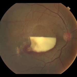
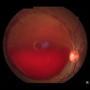 Sub ILM Hemorrhage
Jun 21 2025 by Moazzam Parvez
Fundus photograph of a 46 year old female presenting with a massive sharply demarcated, dome shaped bleed in her right eye.
Photographer: Moazzam Parvez , Netralayam , Kolkata
Imaging device: Topcon Maestro 2
Condition/keywords: sub ILM hemorrhage
-
 Sub ILM Hemorrhage
May 2 2019 by S. Natarajan, MD, FASRS, FRCS (GLASGOW) , FICO, D.Sc, FELA
Fundus photograph of an 56-year-old anemic male who presented with sub ILM hemorrhage at the macula in left eye.
Photographer: Ashwini borde
Imaging device: Carl Zeiss 450 Plus IR
Condition/keywords: hemorrhage, internal limiting membrane (ILM) peeling
-
 Sub ILM Hemorrhage
Jul 29 2014 by Mallika Goyal, MD
Fundus photograph of the left eye of a 26-year-old lady who presented with sudden vision drop (VA 20/40) reveals sub-ILM hemorrhage. There was no history of trauma, valsalva maneuvre or other contributing factors. This heme cleared within 10 days following gas injection and prone positioning with visual recovery to 20/20.
Photographer: Mallika Goyal, MD, Apollo Health City, Jubilee Hills, Hyderabad-500033
Condition/keywords: sub-inner limiting membrane hemorrhage
-
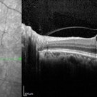
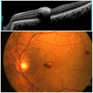
 Sub ILM Hemorrhage
Jul 29 2014 by Mallika Goyal, MD
OCT of the left eye of a 26-year-old lady who presented with sudden vision drop (VA 20/40) reveals sub-ILM haemorrhage. There was no history of trauma, valsalva maneuvre or other contributing factors. This heme cleared within 10 days following gas injection and prone positioning with visual recovery to 20/20.
Photographer: Mallika Goyal, MD, Apollo Health City, Jubilee Hills, Hyderabad-500033
Condition/keywords: sub-inner limiting membrane hemorrhage
-
 Sub ILM Hemorrhage / CPR
Mar 3 2014 by David Callanan, MD
31-year-old female, undergoing hand surgery and suffered cardiac arrest; had CPR in coma for 2 days; noted field defect when she awoke; 20/40 initially 20/20 at end.
Condition/keywords: internal limiting membrane (ILM) peeling
-
 Sub ILM Hemorrhage / CPR
Mar 3 2014 by David Callanan, MD
31-year-old female, undergoing hand surgery and suffered cardiac arrest; had CPR in coma for 2 days; noted field defect when she awoke; 20/40 initially 20/20 at end.
Condition/keywords: internal limiting membrane (ILM) peeling
-
 Sub ILM Hemorrhage / CPR
Mar 3 2014 by David Callanan, MD
31-year-old female, undergoing hand surgery and suffered cardiac arrest; had CPR in coma for 2 days; noted field defect when she awoke; 20/40 initially 20/20 at end.
Condition/keywords: internal limiting membrane (ILM) peeling
 Loading…