 Initializing download.
-
 By Ramses Rosales-Diaz
Asociación Para Evitar la Ceguera en México I.A.P.
Co-author(s): Samuel Peña-Ortiz, Asociación Para Evitar la Ceguera en México - Uploaded on Nov 27, 2024.
- Last modified by Joshua Friedman on Dec 2, 2024.
- Rating
- Appears in
- Miscellaneous
- Condition/keywords
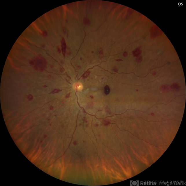
- leukemia, white centered retinal hemorrhage (Roth Spot), sub ILM hemorrhage
- Photographer
- Ramses Rosales-Diaz, Asociación Para Evitar la Ceguera en México
- Imaging device
-
Fundus camera
Clarus 700 - Description
- Fundus photograph of a 48-year-old woman with venous dilatation and tortuosity, flame-shaped and intraretinal hemorrhages, Roth spots and sub-ILM hemorrhage. Her complete blood count reports 425,540 lymphocytes/microliter, and the blood smear reveals Gumprecht shadows and numerous lymphocytes with nuclei exhibiting hypercondensed chromatin. She is diagnosed with chronic lymphocytic leukemia and receives appropriate treatment from the hematology team


---thumb.jpg/image-square;max$79,0.ImageHandler "Roth Spot")
---thumb.jpg/image-square;max$79,0.ImageHandler "Roth Spot")


---thumb.jpg/image-square;max$79,0.ImageHandler "CMV with leukemia")