 Initializing download.
-
 By Ramses Rosales-Diaz
Asociación Para Evitar la Ceguera en México I.A.P.
Co-author(s): Samuel Peña-Ortiz, Asociación Para Evitar la Ceguera en México - Uploaded on Nov 27, 2024.
- Last modified by Joshua Friedman on Dec 2, 2024.
- Rating
- Appears in
- Miscellaneous
- Condition/keywords
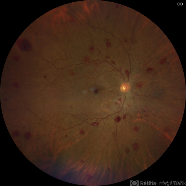
- leukemia, white centered retinal hemorrhage (Roth Spot), sub ILM hemorrhage
- Photographer
- Ramses Rosales-Diaz, Asociación Para Evitar la Ceguera en México
- Imaging device
-
Fundus camera
Zeiss Clarus 700 - Description
- Fundus photograph of a 48-year-old woman showing venous dilatation and tortuosity, flame-shaped hemorrhages, intraretinal hemorrhages, sub-ILM hemorrhages, and Roth spots. Her complete blood count shows 425,540 lymphocytes/microliter, and the blood smear reveals Gumprecht shadows along with numerous lymphocytes with hypercondensed chromatin in their nuclei. She is diagnosed with chronic lymphocytic leukemia and receives appropriate treatment from the hematology team.


---thumb.jpg/image-square;max$79,0.ImageHandler "Roth Spot")
---thumb.jpg/image-square;max$79,0.ImageHandler "Roth Spot")


---thumb.jpg/image-square;max$79,0.ImageHandler "CMV with leukemia")