Search results (141 results)
-
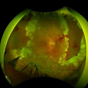
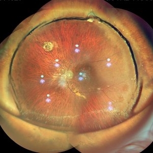 Buckled Silicon oil Filled eye
Jul 25 2019 by Manish Nagpal, MD, FRCS (UK), FASRS
Wide field view of a buckled eye along with silicon oil reflex.
Photographer: Gayathri Mohan, Retina Foundation
Imaging device: Nidek Mirante SLO
Condition/keywords: scleral buckle, silicone oil
-
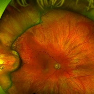
 Choroidal Vessels Transillumination
Mar 20 2024 by Kingston Rodolfo Ureña-Wong, MD, Opht, MSc
Photograph of choroidal vessels transillumination during a scleral buckle to repair a complete retinal detachment.
Photographer: Garagarza-Mariscal Heber, APEC.
Condition/keywords: choroidal vessels, scleral buckle
-
 Erosion of Segmental Buckle
Feb 25 2022 by Roger A. Goldberg, MD, MBA
Erosion of sharp edge of segmental scleral buckle seen 15 years after being placed for repair of a retinal detachment
Photographer: Melissa Bartlett, Bay Area Retina Associates
Imaging device: Optos
Condition/keywords: retinal defect, scleral buckle
-
 MIRAgel Intrusion S/P Scleral Buckle 16 Years Ago
Sep 23 2017 by Timothy S Fuller, MD
Wide-angle fundus photograph of a 75-year-old woman who had a MIRAgel sponge for retinal detachment repair 16+ years ago. The eye has remained comfortable, cosmetically acceptable, and with vision correctable to 20/30. The patient is not bothered by any field defect.
Photographer: Tina Stanley, Texas Retina Associates
Imaging device: Optos
Condition/keywords: scleral buckle
-
 Tractional Detachment of Retina
Aug 21 2024 by Jordyn Beckman
18 year old male with tractional detachment of Retina, chronic macular hole and silicone oil s/p RD repair x2. BCVA CF @2 ft, fellow eye prosthetic.
Photographer: Jordyn Beckman
Imaging device: Optos California
Condition/keywords: Macular hole, preretinal fibrosis, Retinal Detachment, scleral buckle, silicone oil, TRACTION, tractional retinal detachment
-
 Branch Retinal Vein Occlusion
Dec 9 2020 by Olivia Rainey
Ultra-widefield angiogram of a 78-year-old male with a branch retinal vein occlusion affecting his right eye. The patient was diagnosed on 12/17/12 at another practice. The physician noted that there wasn't NVE noted, however areas of micoaneurysmal dilation is present. She noted retinal ischemia secondary to BRVO. 12/8/20 leakage on FA noted to be worsening compared to his previous angiography. She has concern for progressing NVE and recommends sector PRP after injection of antiVEGF series of 3 for the health of the eye.
Photographer: Olivia Rainey, OCT-C, COA
Imaging device: Optos California
Condition/keywords: branch retinal vein occlusion (BRVO), macular branch retinal vein occlusion (BRVO), non-perfusion, scleral buckle, vitreoretinal surgery
-
---thumb.jpg/image-square;max$300,300.ImageHandler) C3F8 gas bubble after retinal detachment surgery
C3F8 gas bubble after retinal detachment surgery
Feb 1 2013 by Sharon Fekrat, MD FACS FASRS
63 year old man s/p encircling scleral buckle and 23g pars plana vitrectomy for a macula off phakic rhegmatogenous retinal detachment. This fundus photograph shows the effect of the encircling buckle and the residual C3F8 intravitreal gas bubble in the right eye.
Photographer: Tiffanie Keaton, Duke Eye Imaging, Duke University Eye Center, Durham, NC
Imaging device: Optos
Condition/keywords: intravitreal gas bubble, vitrectomy
-
 Emulsified Silicone Oil
Apr 3 2025 by Andrew A. Moshfeghi, MD, MBA, FASRS
This is an 87 year- old male with 3.5 year history of retained silicone oil following treatment of late-onset recurrent retinal detachment 18 years following prior primary scleral buckle repair. Robust emulsified silicone oil aggregates are appreciated. Visual acuity is 20/400.
Photographer: Tammy Schoenholz, University of Southern California.
Imaging device: Zeiss Clarus
Condition/keywords: emulsified silicone oil
-
 Exposed Buckle
Dec 28 2012 by Carl C. Awh, MD, FASRS
Pt was referred to Tennessee Retina for an exposed buckle.
Photographer: Alecia Camp, CRA - Tennessee Retina - Nashville, TN
Condition/keywords: exposed scleral buckle
-
 Moyamoya: FA 2 Min OD of an Acute CRAO with CRA Sparing
Nov 17 2019 by John S. King, MD
60-year-old white female presented with five days of acute vision loss in the right eye. She was seen initially by referring doctor after hours five days ago and diagnosed with a CRAO and sent to ED to be evaluated stroke team. Right ICA was 100% closed but completely bypassed. She called four days later c/o redness and eye pain; at this point prominent iris vessels were seen, and she was sent to us. Her background history includes a diagnosis of moyamoya (underwent bilateral cerebral artery bypass 2015); atorvastatin for hypercholesterolemia; ASA; no hx of HTN or heart disease. She had a scleral buckle repair OD in 2017 and later developed a thick ERM, which was repaired in 2018; on her previous visit her acuity was noted at 20/40. On presentation her visual acuity was HM OD and 20/15 OS. IOP was 8 OD and 10 OS. There were prominent iris vessels in the right eye, no cell or flare, and an IOL. The posterior segment exam showed diffuse retinal whitening with attenuated vessels and boxcarring; there was sparing retinal whitening in a central area of the macula that appeared to be supplied by a cilio-retina artery. The FA showed very slow filling of the retinal vessels; there was some early perfusion secondary to the cilio-retinal artery. At 7 minutes there was still significant areas of non-perfusion, as well as macular ischemia. Avastin was administered, and one week later, PRP was performed. On the day PRP was performed, the irregular iris vessels had regressed completely. She said that she had a "sliver" of vision centrally in that eye; her acuity was CF 2' and IOP 12.
Photographer: Gretchen Harper
Imaging device: Topcon
Condition/keywords: central retinal artery occlusion (CRAO), cilioretinal sparing, moyamoya, neovascularization of iris (NVI)
-
 Pseudophakic RRD, S/P Buckle/Vit. w/ Residual Gas Fish Eggs OD
May 23 2018 by Hosam Attia, MD
71-year-old male, s/p combined buckle vitrectomy for recurrent, macula-off, rhegmatogenous retinal detachment, with residual gas fish eggs OD.
Imaging device: Optos California Ultra-Wide Field Fundus Camera
Condition/keywords: encircling scleral buckle, gas bubble, intraocular gas, intravitreal gas bubble
-
 Repaired Retinal Detachment
Jun 24 2025 by Kimberly Wakester
Optomap RGB of an 45-year-old woman with a repaired retinal detachment in the right eye. The operative eye is doing well three-month s/p surgery. Retina is attached 360 on SB. There is resolving residual SRF at 6:00. Discussed the possible need for added laser. Will continue to observe and will return in 3 months for follow up exam.
Photographer: Kimberly Wakester, COA, OCT-C
Imaging device: Optos California
Condition/keywords: repaired RD, scleral buckle
-
 Repaired Retinal Detachment
May 7 2025 by Kimberly Wakester
Optomap RGB montage of an 56-year-old woman with a repaired retinal detachment with scleral buckle and cryotherapy in the left eye. Patient remains stable s/p Vitreo-retinal surgery in 2007. Patient is to return in 1 year for follow up exam with repeat imaging.
Photographer: Kimberly Wakester, COA, OCT-C
Imaging device: Optos California
Condition/keywords: cryotherapy, repaired RD, scleral buckle
-
 Repaired Retinal Detachment with Scleral Buckle
Mar 25 2025 by Kimberly Wakester
Optomap RGB montage of an 64-year-old woman with a repaired retinal detachment with scleral buckle in the right eye. There is nasal and inferior pre-retinal membranes with traction. PPV was recommended but patient defers to proceed with sx at this time. Will continue to follow patient closely for worsening traction. Patient was educated on how to monitor their peripheral vision and was advised to report any changes immediately.
Photographer: Kimberly Wakester, COA, OCT-C
Imaging device: Optos California
Condition/keywords: pre-retinal membrane with traction, repaired RD, scleral buckle
-
 Retinal Detachment Following Scleral Buckling, Retinectomy, Laser, and Oil
Jan 31 2022 by Ahmad B. Tarabishy, MD
Ultra wide-field fundus photograph of a 55-year-old gentleman who is 4 days after surgery with scleral buckling, pars plana vitrectomy, perfluoron tamponade, membrane peeling, direct fluid-PFO-oil exchange, nasal and temporal retinectomies, and endolaser photocoagulation. Visual acuity was 20/150 under oil.
Photographer: Megan McLandsborough, Lakeland Eye Clinic
Imaging device: Optos California UWF Camera
Condition/keywords: endolaser, Membrane Peel, PPV, proliferative retinopathy, proliferative vitreoretinopathy (PVR), Retinal Detachment, retinal detachment with retinal defect, scleral buckle, submacular perfluorocarbon liquid (PFO)
-
---thumb.JPG/image-square;max$300,300.ImageHandler) Retinal Detachment With Dislocated IOL Lens
Retinal Detachment With Dislocated IOL Lens
Jun 30 2013 by Jason S. Calhoun
47-year-old male who had trauma to the right eye. Patient had retinal detachment surgery in the past (scleral buckle), to the right eye. Patient came in with another retinal detachment with dislocated PC IOL lens. Notice the haptics tearing the retina. Patient underwent vitrectomy with gas exchange. VA was hand motion 1 day post-op.
Photographer: Jason S. Calhoun, Mayo Clinic Jacksonville, Florida
Condition/keywords: dislocated posterior chamber intraocular lens (PCIOL), retinal tear
-
 Scleral Buckling IOL Drop
Aug 6 2023 by Dr.Sheetal Divate
A 27 year old female with an old history of trauma and operated with scleral buckling and cataract surgery in the past came recently with complaints of DOV . Findings noted where IOL drop, inferior retinal detachment and old scleral buckle indent.
Photographer: Dr.Sheetal Divate
Imaging device: Optos Advance
Condition/keywords: dislocated intraocular lens (IOL), Retinal Detachment, scleral buckle
-
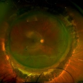
 Attached Retina in a Silicon Oil Filled Buckled Eye with Retinectomy
Apr 17 2021 by Navneet Mehrotra, DNB
Fundus photograph of a 12-year-old boy operated for re retinal detachment with PVR showing attached retina with fresh and old laser marks, silicon oil filled and relaxing retinectomy.
Photographer: Dr Nivesh Gupta, Retina Foundation
Imaging device: Nidek mirante
Condition/keywords: proliferative vitreoretinopathy (PVR), retinectomy, scleral buckle
-
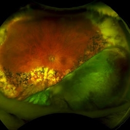
 Total Retinal Detachment
May 27 2020 by Jason Griffith
25-year-old male with histroy of blunt force trauma, s/p RD repair with hx scleral buckle/cryo.
Photographer: Hollie Sanders, Tennessee Retina, Nashville, TN
Imaging device: Optos California
-
 Extruding Buckle
Jul 9 2012 by George W. Aylward, MD, FRCS, FRCOphth
Extruding buckle in a patient who had surgery 5 years previously and re-presented with discomfort and sticky discharge. The buckle was removed following which the retina remained attached, and the symptoms resolved.
Condition/keywords: extruded scleral buckle
-
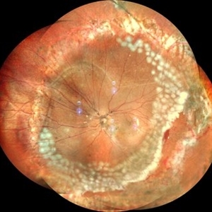
 Vitreous Base Avulsion
Sep 19 2019 by Anfisa Ayalon, MD
Fundus picture of a 34-year-old patient with left eye vitreous base avulsion three months after rhegmatogenous retinal detachment repair with circular scleral buckle implantation. Note bucket handle sign and 360 degrees scleral buckle indentation with a flat retina.
Photographer: Anfisa Ayalon, MD., Meir Medical Center, Kfar Saba, Israel.
Imaging device: California, Optos 200 DTX
Condition/keywords: avulsed vitreous base, behind the vitreous base, scleral buckle
-
 Optos Picture With Speculum: Dislocated Natural Lens
Oct 9 2018 by John S. King, MD
55-year-old white female with history of pathologic myopia+, lattice (laser), SB OU (1990s), and dislocated natural lenses OU that had been watched for years. In the fellow eye she developed phacolytic glaucoma and a PPV, PPL was performed. Plan for both eyes are monitoring. I wanted to get a good picture of her lens today with the optos machine, as the other pics had artifact from the lower lid. It worked out well to use a speculum in the left eye. Vision cc is 20/400 J1+ OD and 20/40 J2 OS; aphakic OU; vitreous clear OD; dislocated lens OS (see pic); retinas attached.
Photographer: Maisee Yang
Imaging device: Optos California
Condition/keywords: dislocated crystalline lens, pathologic myopia, scleral buckle, staphyloma
-
 UWF of Retinal Detachment Corrected with Scleral Buckle
Aug 29 2017 by Carolyn Daley
An ultra wide field fundus photograph of a 57-year-old male who has a past history of retinal detachment corrected with scleral buckle and three treated retinal tears.
Photographer: Carolyn Daley
Imaging device: Optos Imaging
Condition/keywords: cryo-retinal tear, cryotherapy, Optos, retinal tear, scleral buckle, ultra-wide field imaging
-
 Pseudophakic RRD, S/P Buckle/Vit. w/ Residual Gas Fish Eggs OD
May 23 2018 by Hosam Attia, MD
71-year-old male, s/p combined buckle vitrectomy for recurrent, macula-off, rhegmatogenous retinal detachment, with residual gas fish eggs OD.
Imaging device: Optos California Ultra-Wide Field Fundus Camera
Condition/keywords: encircling scleral buckle, gas bubble, intraocular gas, intravitreal gas bubble
-
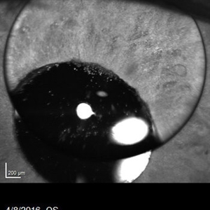 Silicone Oil Bubble in Anterior Chamber - 15 Degree Angle
Apr 11 2016 by Zach Dupureur
30 % silicone oil bubble involving central visual axis. Occurred after a PPV with silicone oil. Oil from vitreous moved into the anterior chamber.
Photographer: Zachary Dupureur, OCT-C
Imaging device: Heidelberg Spectralis
Condition/keywords: anterior chamber, detachment, infrared image, pars plana vitrectomy (PPV), scleral buckle, silicone oil
 Loading…