 Initializing download.
-
 By JEFFERSON R SOUSA, Tecg.º (Biomedical Systems Technology)
Lens Oftalmologia - Hospital Beneficiência Portuguesa - Uploaded on Mar 28, 2021.
- Last modified by JEFFERSON R SOUSA, Tecg.º (Biomedical Systems Technology) on Nov 9, 2021.
- Rating
- Appears in
- Miscellaneous
- Condition/keywords
- retinoschisis, Coats' disease
- Photographer
- JEFFERSON R SOUSA - Study Center and Ophthalmological Research Dr. Andre M V Gomes, Institute Dr. Suel Abujamra São Paulo-Brazil
- Imaging device
-
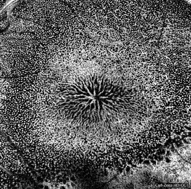
Optical coherence tomography system
Optical coherence tomography system Optical Coherence Tomography system OCT CIRRUS 5000, Line Protocol, HD 21 line. Cirrus 5000 does not do a wide-angle tomographic image. (Structrure Deep: IPL - 25, OPL - 25). - Description
- A 14-year-old male patient was admitted for visual assessment. Visual acuity s/c in the right eye and 20/80 in the left eye. According to family members, he reported low vision since childhood. He had already undergone photocoagulation treatment at another service for which he had a diagnostic hypothesis of Coats' disease. Laboratory tests were requested (HIV, TOXO, TOXOCARIASIS, ACE, VDRL, PPD). In the evaluation, there was significant exudation in the posterior pole, some vascular irregularities in the right eye. In the left eye, there is retinoschisis affecting the entire posterior pole and the nasal region to the optic disc, macula with a characteristic chariot-wheel appearance, well exemplified by OCT-A (Structrure Deep: IPL - 25, OPL - 25).


")


---thumb.jpg/image-square;max$79,0.ImageHandler "Retinoschisis")


