Search results (150 results)
-
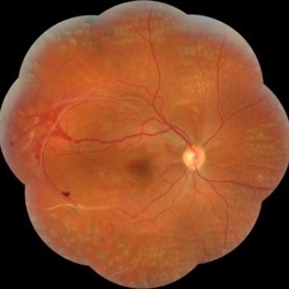
 Proliferative Diabetic Retinopathy with Pre-retinal Hemorrhage
Jan 16 2018 by Olivia Rainey
Ultra-wide field pseudo-color image of an 57-year-old male with a large pre-retinal hemorrhage secondary to proliferative diabetic retinopathy affecting his left eye.
Photographer: Olivia Rainey
Imaging device: Optos California
Condition/keywords: color fundus photograph, diabetic mellitus, hemorrhage, left eye, neovascularization (NV), Optos, proliferative diabetic retinopathy (PDR), pseudocolor, ultra-wide field imaging
-
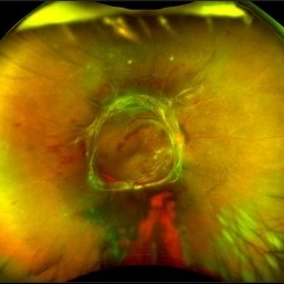
 Proliferative Sickle Cell Retinopathy
Feb 1 2023 by Olivia Rainey
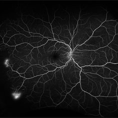
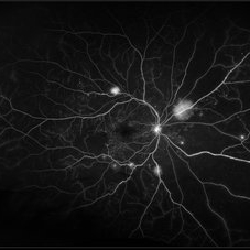
Ultra-widefield fluorescein angiography of a 25-year old male with Proliferative Sickle Cell Retinopathy affecting his right eye. Patient stated that he was born with Sickle disease (SC), and has yearly eye exams. He noted no vision concerns over the last year but has typically experienced sickle attacks about 1-2 per year. The physician noted that the fluorescein obtained showed peripheral nonperfusion affecting the patient's nasal and temporal retina as well as neovascularization affecting his left eye more than his right. He recommended pan retinal photocoagulation in his left eye for his temporal and nasal retina, as as well as his right eye following.
Photographer: Olivia Rainey, OCT-C, COA
Imaging device: Optos California
Condition/keywords: early phase, fluorescein angiogram (FA), fluorescein leakage, neovascularization (NV), non-perfusion, proliferative retinopathy, right eye, sickle cell retinopathy, ultra-wide field imaging, ultra-widefield image
-
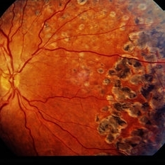
 Sickle Cell Retinopathy
Feb 15 2021 by Kim Barrett
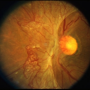
24-year-old female with Sickle Cell Retinopathy, stage 3. She confirms she has the trait as well as her grandmother, mother and a sibling. She has seafan neovascularization superotemporal OD. Current VA is 20/20. Photo is pre-PRP laser with areas of non-profusion temporally.
Photographer: Kim Barrett C.O.A. Retina Specialist of Michigan, Grand Rapids, MI
Imaging device: Optos California
Condition/keywords: neovascularization (NV), pan-retinal photocoagulation (PRP), sickle cell retinopathy, stage 3, trait
-
 Advanced Active PDR
Mar 29 2013 by Henry J. Kaplan, MD
Extensive NVD-FPD and NVE-FPE in a diabetic patient.
Condition/keywords: foveal photoreceptor defect, FPE, neovascularization (NV), neovascularization of the disc (NVD)
-
 Diabetic Macular Edema, Proliferative Diabetic Retinopathy, Neovascularization Elsewhere, DME, PDR, NVE
Apr 1 2013 by James B. Soque, CRA, OCT-C, COA, FOPS
39-year-old white female and long standing diabetis, c/o new peripheral symptoms of left eye. FA OS reveals diabetic macular edema, microaneurysms, and neovasculaization elsewhere. Fluorescein Angogram, Early Phase, 50 Deg, 2x Mag.
Photographer: James B Soque, CRA, COA
Imaging device: Topcon TRC 50DX with MERGE software, OIS 10.6.45
Condition/keywords: diabetic macular edema, neovascularization (NV), proliferative diabetic retinopathy (PDR)
-
 Extensive Pan-Retinal Photocoagulation
Apr 19 2013 by Brandon G. Busbee, MD
Extensive pan-retinal photocoagulation.
Photographer: Alecia Camp, CRA - Tennessee Retina - Nashville, TN
Imaging device: Topcon TRC 50-EX
Condition/keywords: neovascularization (NV), pan-retinal photocoagulation (PRP)
-
 Neovascularization of the Disc
Oct 27 2017 by Claire Kiernan, MD
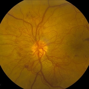
Optic disc photograph of a 53-year-old diabetic man with new optic disc neovascularization on dilated eye exam.
Photographer: Joseph Mastellone, Steve Moser
Condition/keywords: diabetic mellitus, neovascularization (NV), neovascularization of the disc (NVD)
-
 Proliferative Diabetic Retinopathy
May 15 2018 by Morgan Benton
Ultra-wide field pseudocolor image of a 42-year-old female with proliferative diabetic retinopathy resulting in severe hemorrhaging. Vision was cc20/80+1 when the image was taken.
Photographer: Morgan Benton
Imaging device: Optos
Condition/keywords: color fundus photograph, hemorrhage, left eye, montage, neovascularization (NV), Optos, proliferative diabetic retinopathy (PDR), ultra-wide field imaging
-
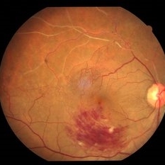
 Proliferative Diabetic Retinopathy
May 11 2020 by Gayathri Mohan
Color fundus photograph of a patient with PDR, showing neovascularisation infero-temporal to macula.
Photographer: Gayathri Mohan, Retina Foundation
Imaging device: Mirante, Nidek
Condition/keywords: neovascularization (NV), optical coherence tomography (OCT), proliferative diabetic retinopathy (PDR)
-
 Proliferative Diabetic Retinopathy
Jan 29 2021 by Olivia Rainey
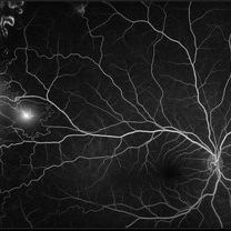
Ultra-widefield fluorescein angiogram of a 65-year-old male with proliferative diabetic retinopathy affecting his right eye. The patient's diabetic retinopathy has progressed significantly since he was last seen in 2014. It was recommended to begin antiVEGF to control DME followed by laser treatment OU.
Photographer: Olivia Rainey, OCT-C, COA
Imaging device: Optos California
Condition/keywords: anti-VEGF, diabetes, diabetic macular edema, fluorescein angiogram (FA), fluorescein leakage, neovascularization (NV), neovascularization elsewhere (NVE), non-perfusion, Optos, proliferative diabetic retinopathy (PDR), ultra-wide field imaging
-
 Proliferative Diabetic Retinopathy
Mar 16 2015 by Matt Poe, COA
IVFA of 53-year-old male with Proliferative Diabetic Retinopathy, Diabetic macular edema, and a tractional retinal detachment.
Photographer: Matt Poe, COA. Northwest Arkansas Retina Associates, Springdale, AR.
Imaging device: Heidelberg HRA
Condition/keywords: neovascularization (NV), proliferative diabetic retinopathy (PDR)
-
---thumb.jpg/image-square;max$300,300.ImageHandler) Proliferative Diabetic Retinopathy (PDR) & Traction Retinal Detachment
Proliferative Diabetic Retinopathy (PDR) & Traction Retinal Detachment
Feb 13 2013 by From the Collections of Thomas M. Aaberg, MD and Thomas M. Aaberg Jr., MD
Florid NV with early macular TRD.
Condition/keywords: neovascularization (NV), tractional retinal detachment
-
 Proliferative Sickle Cell Retinopathy
Jan 29 2021 by Olivia Rainey
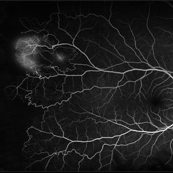
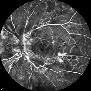
Ultra-widefield fluorescein angiogram of a 24-year-old female with proliferative sickle cell retinopathy affecting her right eye. The physician's interpretation of the fluorescein shows seafan neovascularization superotemporally, AV anastomeses, and good peripheral laser. He performed scatter PRP OD on 12/2/2020 to nonperfusion in temporal far periphery. The patient's 12/2020 Hb electrophoresis came back showing Hb SC (rather than sickle cell trait). Patient was born at full term, but she reports that her mother used drugs while pregnant with the patient. The patient also mentioned that her niece has full sickle cell disease and her grandmother, mother, and sibling all have condition on the sickle cell spectrum.
Photographer: Olivia Rainey, OCT-C, COA
Imaging device: Optos California
Condition/keywords: fluorescein angiogram (FA), fluorescein leakage, neovascularization (NV), neovascularization elsewhere (NVE), Optos, sea fan, sickle cell retinopathy
-
 Severe NVD
Mar 26 2018 by Kristen Wagner
Fundus photograph of a young woman with uncontrolled Diabetes Type II with severe neovascularization of the disc (NVD) and PDR.
Photographer: Kristen Wagner, COT, OSC
Condition/keywords: diabetes, neovascularization (NV), neovascularization of the disc (NVD), optic disc, optic nerve, proliferative diabetic retinopathy (PDR)
-
 Severe Neovascularization Secondary to Idiopathic Occlusive Retinal Vasculitis
Jan 17 2015 by Hamid Ahmadieh, MD
Wide- field color fundus photograph of the right eye of a 28-year-old woman with severe retinal neovascularization secondary to idiopatic occlusive retinal vasculitis.
Photographer: Solmaz Shahmohammad, Negah Eye Center, Tehran
Condition/keywords: color fundus photograph, neovascularization (NV), retinal vasculitis
-
 Proliferative Diabetic Retinopathy
May 15 2018 by Morgan Benton
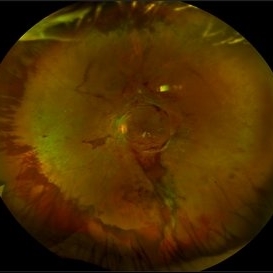
Ultra-wide field pseudocolor image of a 42-year-old female with proliferative diabetic retinopathy resulting in a tractional retinal detachment. Vision was cc20/50-2+1 when the image was taken.
Photographer: Morgan Benton
Imaging device: Optos
Condition/keywords: color fundus photograph, neovascularization (NV), Optos, proliferative diabetic retinopathy (PDR), tractional retinal detachment, ultra-wide field imaging
-
 Sickle Cell Neovascularization and Vitreous Hemorrhage
Oct 30 2015 by David Callanan, MD
Female patient, sickle cell neovascularization and vitreous hemorrhage; pre and post laser.
Condition/keywords: neovascularization (NV), sickle cell, vitreous hemorrhage
-
 Battle of BRVOs: Old vs New
Jul 30 2021 by Gayathri Mohan
Color fundus photograph of a 48-year-old showing an inferno temporal BRVO. Old BRVO with neovascularization seen super-temporally.
Photographer: Dr. Gayathri Mohan
Imaging device: Canon
Condition/keywords: branch retinal vein occlusion (BRVO), cystoid macular edema (CME), neovascularization (NV)
-
 Ischemic BRVO with Neovascularization
Aug 23 2012 by Gerardo Garcia-Aguirre, MD
Fluorescein angiogram of the macula showing wide areas of capillary nonperfusion and leakage in the superotemporal quadrant.
Photographer: Noemí Hernández, Asociación para Evitar la Ceguera en México
Condition/keywords: branch retinal vein occlusion (BRVO), capillary nonperfusion, neovascularization (NV)
-
 Ischemic BRVO with neovascularization
Aug 23 2012 by Gerardo Garcia-Aguirre, MD
Fluorescein angiogram of the temporal periphery showing wide areas of capillary nonperfusion and leakage secondary to neovascularization.
Photographer: Noemí Hernández, Asociación para Evitar la Ceguera en México
Condition/keywords: branch retinal vein occlusion (BRVO), capillary nonperfusion, neovascularization (NV)
-
 Proliferative Diabetic Retinopathy
Aug 23 2012 by Gerardo Garcia-Aguirre, MD
Fluorescein angiogram of a left eye of a 45 year-old patient with proliferative diabetic retinopathy. Small hyperfluorescent dots are observed (microaneurysms), as well as blockage from a subhyaloid hemorrhage. In the inferonasal area two areas of leakage secondary to neovascularization are observed.
Photographer: Noemí Hernández, Asociación para Evitar la Ceguera en México
Condition/keywords: microaneurysms, neovascularization (NV), subhyaloid hemorrhage
-
 Proliferative Diabetic Retinopathy (PDR)
Jul 4 2018 by Deepak Bhojwani, MS
Colour Fundus Photograph of a 66-year-old diabetic male with large fibro-vascular proliferative vessels causing subhayolid haemorrhage and tractional retinal detachment involving posterior pole.
Photographer: Deepak Bhojwani
Condition/keywords: diabetes, neovascularization (NV), subhyaloid hemorrhage, tractional retinal detachment
-
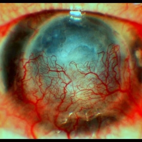 CORNEA
Feb 23 2018 by JEFFERSON R SOUSA, Tecg.º (Biomedical Systems Technology)
64-year-old patient, with vision loss more than 10 years after having suffered blunt trauma with ocular perforation.
Photographer: JEFFERSON R SOUSA - Study Center and Ophthalmological Research Dr. Andre M V Gomes, Institute Dr. Suel Abujamra São Paulo-Brazil
Imaging device: Topcon TRC-50 DX, Imaginet 5.0, angle de 20 graus. Flash 36.
Condition/keywords: 20 degrees, central opacity of cornea, corneal edema, neovascularization (NV)
-
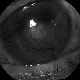 Neovascularization of The Cornea
May 13 2016 by Nichole Lewis
Neovascularization of the Cornea
Photographer: Nichole Lewis
Condition/keywords: cornea, neovascularization (NV)
-
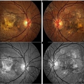 Angioid Streaks
May 11 2016 by Andrea Arriola-Lopez, MD MSc
64-year-old man, VA CF AO. Inactive neovascularization. Color fundus and red free photograph.
Photographer: Andrea E. Arriola-Lopez MD MSc
Imaging device: Visucam lite Zeiss
Condition/keywords: angioid streaks, color fundus photograph, neovascularization (NV), red-free
 Loading…