File number: 28211
Comments
-
 Suber S. Huang, MD, MBA, FASRS (June 7 2018)
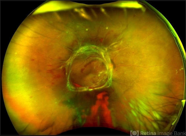
Nice capture! A traditional 55 degree image might give better clinical details. The periphery shows non-perfusion and lashes. A less magnified image without pseudocoloration might better illustrate this end-stage pathology. Thanks for sharing!
Sign in to comment.
 Initializing download.
-
 By Morgan Benton
- Uploaded on May 15, 2018.
- Last modified by Caroline Bozell on May 15, 2018.
- Rating
- Appears in
- Miscellaneous
- Condition/keywords
- proliferative diabetic retinopathy (PDR), tractional retinal detachment, neovascularization (NV), Optos, ultra-wide field imaging, color fundus photograph
- Photographer
- Morgan Benton
- Imaging device
-
Fundus camera
Optos - Description
- Ultra-wide field pseudocolor image of a 42-year-old female with proliferative diabetic retinopathy resulting in a tractional retinal detachment. Vision was cc20/50-2+1 when the image was taken.


