Search results (450 results)
-
 Dropped IOL
Apr 5 2018 by Mohamed Tawfik, MD
Intra operative photo of a case of dropped IOL after phaco+IOL.
Photographer: Mohamed A,Tawfik
Imaging device: Intra operative Photography Screen shoot
Condition/keywords: dropped intraocular lens (IOL)
-
 Pigmented Paravenous Retinochoroidal Atrophy (PPRCA)
Jun 30 2025 by Maria Letícia Costa Holanda
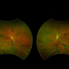
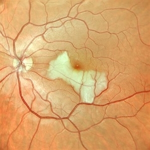
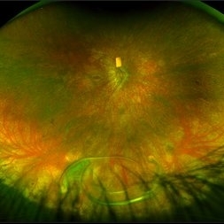
Fundoscopy of a 42-year-old asymptomatic man with pigmented paravenous chorioretinal atrophy. Pigmented paravenous retinochoroidal atrophy (PPRCA) is a rare disorder of unknown etiology. The disease is characterized by pigment accumulation along the distribution of retinal veins. The findings are usually incidental with minimal effect on vision.
Photographer: Guilherme da Cruz Reis, CLINOS Eye Hospital - Feira de Santana (BA),Brazil
Condition/keywords: pigmented paravenous chorioretinal atrophy (PPCRA)
-
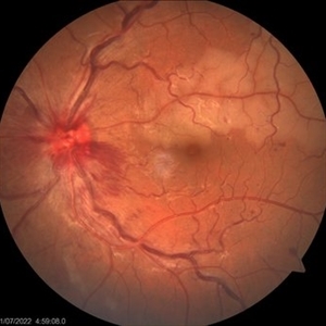 Combined central retinal vein occlusion and branch retinal arteriolar occlusion
Sep 13 2022 by Ruchir Mehta, DO, DNB, FRCS
Fundus photograph of left eye of a 63 years old female with known type 2 DM and HTN showing combined central retinal venous occlusion and superior branch retinal arteriolar occlusion
Photographer: Ruchir Mehta, Mehta Superspeciality Eye Hospital, Jamnagar, Gujarat, India
Imaging device: Zeiss Visucam 500
Condition/keywords: branch retinal artery occlusion (BRAO), central retinal vein occlusion (CRVO), COMBINED
-
 Dislocated IOL
Sep 28 2024 by Anjana Mirajkar, MS Ophthalmology
An intra operative image of right eye showing dislocated IOL sitting on the posterior pole.
Photographer: Dr. Anjana Mirajkar -Retina Foundation, Ahmedabad
Condition/keywords: dislocated IOL
-
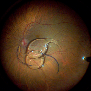 Fraternal Twins
May 22 2023 by Gustavo M. Hüning, MD, MBA, FASRS
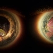
Intrasurgical photograph using a non-contact system and 3D visualization system of a 65-year-old woman who suffered an ocular trauma.
Photographer: Gustavo M. Hüning, Hüning Clínica do Olhar, Santa Maria - Brazil
Imaging device: Alcon Luxor combined with Alcon nGenuity
Condition/keywords: dislocated intraocular lens (IOL), implant, pars plana vitrectomy (PPV)
-
 Pigmented Paravenous Chorioretinal Atrophy (PPCRA)
Jun 27 2025 by Maria Letícia Costa Holanda
Fundoscopy of a 42-year-old asymptomatic man with pigmented paravenous chorioretinal atrophy. Pigmented paravenous retinochoroidal atrophy (PPRCA) is a rare disorder of unknown etiology. The disease is characterized by pigment accumulation along the distribution of retinal veins. The findings are usually incidental with minimal effect on vision.
Photographer: Guilherme da Cruz Reis, CLINOS Eye Hospital - Feira de Santana (BA),Brazil
Condition/keywords: pigmented paravenous chorioretinal atrophy (PPCRA)
-
 Pigmented Paravenous Retinochoroidal Atrophy (PPRCA)
Jun 27 2025 by Maria Letícia Costa Holanda
Fundoscopy of a 42-year-old asymptomatic man with pigmented paravenous chorioretinal atrophy. Pigmented paravenous retinochoroidal atrophy (PPRCA) is a rare disorder of unknown etiology. The disease is characterized by pigment accumulation along the distribution of retinal veins. The findings are usually incidental with minimal effect on vision.
Photographer: Guilherme da Cruz Reis, CLINOS Eye Hospital - Feira de Santana (BA),Brazil
Condition/keywords: pigmented paravenous chorioretinal atrophy (PPCRA)
-
 Rescuing IOL CTR Bag Complex
Jun 14 2023 by Aditya S Kelkar, MS, FRCS, FASRS,FRCOphth
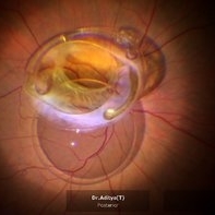
INTRAOPERATIVE SNAPSHOPT IN ZEISS ARTEVO 800 OF DROPPED IOL CTR BAG COMPLEX IN A 71 YEAR OLD MALE PATIENT
Photographer: SUBHASREE DUTTA, NATIONAL INSTITUTE OF OPHTHALMOLOGY, PUNE
Imaging device: ZEISS ARTEVO 800
Condition/keywords: dropped capsular IOL bag complex
-
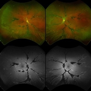
 Retinitis Pigmentosa with PPRPE
Jan 27 2025 by Vishal Agrawal, MD, FRCS,FACS,FASRS
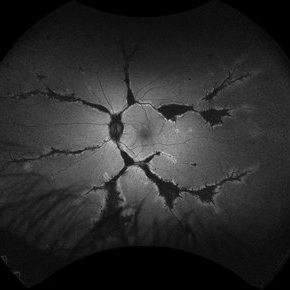
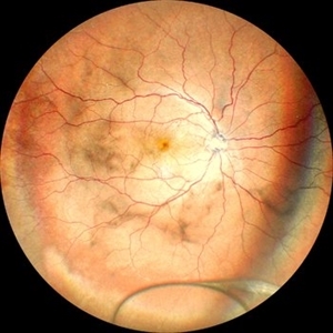
16 year-old male patient presented with DOV, nyctalopia and nystagmus. Fundus revealed pigment clumping, pale disc and preserved para-arteriolar retinal pigment epithelium (PPRPE) in both eyes. Genetic testing revealed CRB1 gene mutation.
Photographer: Dr Ayushi
Imaging device: Clarus 700
Condition/keywords: retinitis pigmentosa
-
 Valsalva retinopathy progression
Oct 4 2023 by Niloofar Piri, MD
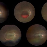
Progression fundus photograph images of a 22 yo female with Valsalva retinopathy secondary to violent emesis. Note the sub ILM layered hemorrhage and gradual decrease then disappearance over time. Last image is at 6 month follow up with 20/20 vision. Of note the silhouette of ILM separation is still visible.
Condition/keywords: valsalva retinopathy
-
 Arteriolar Macruaneurysm
Jul 11 2013 by Jerald A. Bovino, MD
No history, with hemorrhage.
Condition/keywords: arteriolar macroaneurysm
-
 Central Retinal Artery Occlusion
Jan 22 2021 by Renata Garcia Franco, Md
65-year-old male, history of uncontrolled systemic arterial hypertension. Segmentation of blood in retinal arterioles, retinal whitening and cherry red spot.
Photographer: Fatima Hernandez, Instituto de la Retina del Bajio SC
Imaging device: Zeiss
Condition/keywords: central retinal artery occlusion (CRAO)
-
 Cilioretinal Artery Occlusion
May 14 2024 by Eloy Mata-Cortes, MD
Color image capturing the left eye of a 32-year-old female. Despite a negative ophthalmological and medical history, she reported three days of blurred vision and a paracentral scotoma in her left eye, while maintaining central vision. The image reveals retinal whitening, extends from the parafoveal region to the inferotemporal arcade indicative of cilioretinal artery occlusion. Following this observation, the patient was referred for systemic assessment to explore the underlying etiology of the occlusion.
Photographer: Eloy Mata-Cortes, MD, Instituto Mexicano de Oftalmología, Querétaro, México
Imaging device: Nidek Mirante
Condition/keywords: cilioretinal artery occlusion, oclussion, retinal whitening
-
 CRVO with Secondary CLRAO
May 28 2020 by Richard M Martindale, MD
Non-ischemic CRVO (VA 20/30) with secondary CLRAO (nasal macular pallor) in a hypertensive 69yo female. Pathophysiologically, the cilioretinal artery occlusion occurs secondary to elevation in the hydrostatic pressure in the retinal venous system relative to the choroidal perfusion pressure (which supplies the cilioretinal artery).
Photographer: Retina Consultants of Alabama, P. C.
Imaging device: Optos
Condition/keywords: cilioretinal artery occlusion, non-ischemic central retinal vein occlusion (CRVO)
-
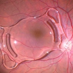
 Dislocated Crystalline Lens
Mar 19 2024 by Annaka Gooding
Ultra Wide field fundus photography of a 70 year old male who presented to clinic with a sudden increase of vision due to dropped crystalline lens secondary to severely dense cataract. Patient reported seeing a full black circle in his inferior visual field. Patient's visual acuity at time of visit was 20/100 with a +5.00 diopter lens. The physician recommended surgical intervention, and discussed surgery for PPV/PPL/IOL implantation with an ACIOL.
Photographer: Annaka Gooding, CPO
Imaging device: Optos California RGB
Condition/keywords: dislocated crystalline lens, fundus photography, inferior retina, OPTOS CALIFORNIA RGB, Right Eye, Ultra-wide field retinal imaging
-
 Dislocated Intraocular Lens
Nov 15 2024 by Tejaswita Verma
Fundus image of a spontaneously posteriorly dislocated IOL 10 years following surgery. Other eye had a subluxated opacified IOL.
Photographer: DR. TEJASWITA VERMA
Imaging device: MIRANTE
Condition/keywords: dislocated intraocular lens (IOL)
-
 Dislocated Intraocular Lens
Jun 4 2025 by Aditya S Kelkar, MS, FRCS, FASRS,FRCOphth
Fundus photograph of a 79-year-old man with a posteriorly dislocated intraocular lens in the inferior quadrant.
Photographer: Optom Chandrakanta Bhandare, National Institute of Ophthalmology, Pune
Imaging device: Optos Daytona
Condition/keywords: dislocated intraocular lens (IOL)
-
 Dislocated Intraocular Lens (IOL)
Aug 2 2019 by JEFFERSON R SOUSA, Tecg.º (Biomedical Systems Technology)
A 53-year-old male patient suffered blunt trauma 15 days after cataract surgery. Note total dislocation of the intraocular lens. No glass reaction.
Photographer: JEFFERSON R SOUSA - Study Center and Ophthalmological Research Dr. Andre M V Gomes, Institute Dr. Suel Abujamra São Paulo-Brazil
Imaging device: Topcon TRC-50 DX, Imaginet 4.0, angle de 50 graus. Flash 18w-s
Condition/keywords: dislocated intraocular lens (IOL)
-
 Dislocated IOL
May 15 2018 by Morgan Benton
Ultra-wide field pseudocolor image of a 68-year-old male with a dislocated IOL after cataract surgery in the left eye. Patient was only able to count fingers at one foot and could pinhole to 20/60.
Photographer: Morgan Benton
Imaging device: Optos
Condition/keywords: color fundus photograph, dislocated intraocular lens (IOL), left eye, Optos, ultra-wide field imaging
-
 Dislocated IOL
Jan 13 2014 by David Callanan, MD
Dislocated IOL, 63-year-old female.
Condition/keywords: dislocated intraocular lens (IOL)
-
 Dislocated IOL
Jun 4 2024 by Marlee Curnutt
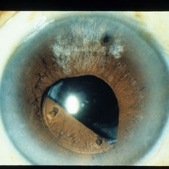
Slit lamp photo of a 64 year old woman presenting with worsening vision and depth perception after trauma induced by a dog, which dislocated her IOL. The patient's IOL haptic was seen in the AC, and almost abutting cornea. Patient's vision upon presentation was DCC CF@1 feet. Patient was counseled and underwent an IOL exchange.
Photographer: Marlee Curnutt, COA
Imaging device: Galaxy A42
Condition/keywords: dislocated intraocular lens (IOL), haptic, IOL, right eye, slit lamp photo, slit lamp photography, trauma
-
 Dislocated Lens
Apr 26 2023 by Chloe Hanifan
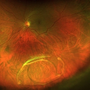
Ultra wide field fundus photograph of a 41-year-old male with a dislocated lens affecting his right eye. IOL noted inferior vitreous base and vitrectomy surgery for removal of IOL was recommended. Patient has history of retinitis pigmentosa as well. Patient's vision at the time of presentation was counting fingers at 2 feet.
Photographer: Chloe Hanifan
Imaging device: Optos California
Condition/keywords: dislocated lens, fundus photography, Optos, pseudocolor, retinitis pigmentosa, ULTRA WIDE FIELD
-
 Dislocated Lens
Jun 29 2013 by Jason S. Calhoun
84-year-old female comes in with blurred vision in the left eye. VA was 20/30, right eye and count fingers in the left eye. Fundus examination reveals dislocation of the IOL into the vitreous inferiorily at 6-o'clock. Suggest surgery to fix the problem.
Photographer: Jason S. Calhoun, Mayo Clinic Jacksonville, Florida
Imaging device: TOPCON TRC 50-EX
Condition/keywords: dislocated posterior chamber intraocular lens (PCIOL)
-
 Dislocated Lens, Posterior OD
Jan 26 2024 by Corey Grant
OPTOS California photo presents a 71 year old male patient with a dislocated lens, posterior in the right eye. Presented on 1/26/24 with posteriorly dislocated SN60WF with a Soemmerring ring. Associated retinal hemorrhage within retinoschisis as well. This will result in a PPV/IOL exchange/SFIOL/STK for the right eye.
Photographer: Corey Grant, Ophthalmic Imager, Retina Specialist of Michigan
Imaging device: OPTOS California
Condition/keywords: color photo, IOL, OD, Optos, OPTOS CALIFORNIA, pars plana vitrectomy (PPV), retina
-
 Dislocated-P/C IOL Bag Complex
Nov 27 2018 by Maria H. Berrocal, MD
85-year-old who underwent phaco IOL 15 years prior, who noticed loss of vision OS.
Photographer: Thaylan Calderon, Berrocal & Associates, San Juan, PR
Imaging device: Optos
Condition/keywords: dislocated intraocular lens (IOL)
 Loading…