Search results (450 results)
-
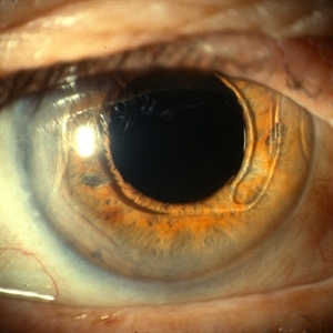 Anterior Chamber Intraocular Lens
Sep 20 2012 by Jeffrey G. Gross, MD, FASRS
AC-IOL, s/p PPV, lensectomy for dislocated crystalline lens, 20/20
Condition/keywords: anterior chamber, dislocated crystalline lens, intraocular lens (IOL), lensectomy
-
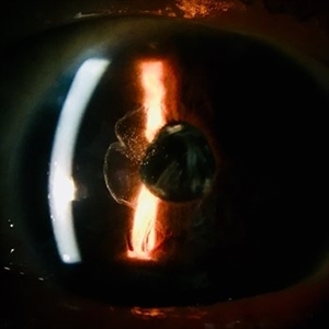
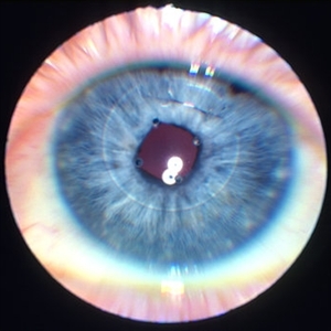 Iris clip IOL
Jan 11 2013 by Alex P. Hunyor, MD
Iris fixated IOL - note stainless steel suture.
Condition/keywords: intraocular lens (IOL), iris clip intraocular lens
-
 Ocular Albinism Carrier
Feb 13 2013 by From the Collections of Thomas M. Aaberg, MD and Thomas M. Aaberg Jr., MD
RPE atrophy, degeneration, thinned arterioles.
Condition/keywords: degeneration, ocular albinism, retinal pigment epithelium atrophy, thinned arterioles
-
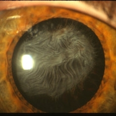 Anterior Capsule Opacification in Eye
Oct 11 2012 by Jeffrey G. Gross, MD, FASRS
Anterior capsule opacification in eye, s/p PPV lensectomy, without IOL.
Condition/keywords: anterior capsule opacification, lensectomy, without intraocular lens
-
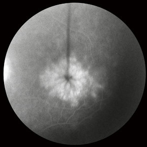
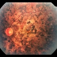 Retinitis Pigmentosa
Apr 6 2022 by Marianna Kavalaraki, MD, Msc
Fundus photography of a 21-year-old man with retinitis pigmentosa. Fundus findings include retinal pigmentary changes in the form of widespread pigment clumpings predominantly in the mid-peripheral fundus, arteriolar attenuation, RPE and retinal atrophy in the posterior pole.
Photographer: Marianna Kavalaraki, General Hospital of Nikaia Piraeus, Department of Ophthalmology
Imaging device: Canon CF-60DSi Digital Fundus Camera
Condition/keywords: retinitis pigmentosa
-
 Ocular Albinism Carrier
Feb 13 2013 by From the Collections of Thomas M. Aaberg, MD and Thomas M. Aaberg Jr., MD
RPE atrophy, degeneration, thinned arterioles.
Condition/keywords: degeneration, ocular albinism, retinal pigment epithelium atrophy, thinned arterioles
-
 Retinal Detachment Repair With Silicone Oil and Scleral Buckle, Fourteen Years Later, With Visual Acuity of 20/25
Sep 12 2016 by Timothy S Fuller, MD
65-year-old woman s/p scleral buckle 14 years ago. Two weeks later, the retina re-detached, and vitrectomy, laser, and silicone oil procedure was performed. Patient remains 20/25 with correction fourteen years later. The cornea is clear, there is no oil emulsification, and there is a stable, moderately inferiorly subluxated PCIOL (as it was prior to RD surgery). IOP is 17 on Cosopt BID.
Photographer: Nicholas Hesse, Texas Retina Associates
Imaging device: Optos
Condition/keywords: laser, scleral buckle, silicone oil
-
 Lattice Degeneration
Nov 9 2012 by Norman Byer
This is a very subtle example of lattice degeneration showing the mildest possible changes in a 27-year-old man. In the upper left there is a vein directed toward the center of the slide. Just above and to the right of the pigment spot it veers to the right and then abruptly disappears as it passes through the lattice lesion. As it leaves the lesion, it resumes its normal appearance going down to the right. In a similar manner, the arteriole in the lower left enters the lesion just to the right of the pigment spot, then disappears as it passes through the lesion and reappears later as it emerges. The only change in this lesion in 12 years was the appearance of the pigment spot.
Condition/keywords: lattice degeneration
-
 Toxoplasmosis Slide 2
Oct 22 2012 by Ronald C. Gentile, MD
One month following treatment with Bactrim, Clindamycin, and oral prednisone the focal area chorioretinitis has coalesced with a decrease in overlying vitreous inflammation. Kyrieleis plaques can be seen along the inferior retinal arteriole.
Photographer: The New York Eye & Ear Infirmary Department of Medical Imaging
Condition/keywords: posterior uveitis, toxoplasmosis
-
 Dislocated IOL With PI
Jul 14 2013 by Jason S. Calhoun
Subluxated lens with PI at 12-o'clock.
Photographer: Jason S. Calhoun, Department of Ophthalmology, Mayo Clinic Jacksonville, Florida
Imaging device: TOPCON D-90 SL NIKON CAMERA
Condition/keywords: dislocated posterior chamber intraocular lens (PCIOL), peripheral iridotomy
-
000---thumb.jpg/image-square;max$300,300.ImageHandler) Dropped IOL into the Vitreous Cavity
Dropped IOL into the Vitreous Cavity
Oct 7 2012 by Young Hee Yoon, MD, PhD
Fundus photograph of an 70-year-old man with a history of cataract operation 20 years ago. He visited our clinic with decreased visual acuity for 2 days.
Photographer: Yoon-hwa Kim, Asan Medical Center
Imaging device: Optomap, optos
Condition/keywords: intraocular lens dislocation
-
 Arteriolar Macroaneurysm
Oct 1 2012 by Jeffrey G. Gross, MD, FASRS
Arteriolar macroaneurysm with partially reabsorbed subretinal hemorrhage.
Condition/keywords: arteriolar macroaneurysm, partially reabsorbed subretinal hemorrhage
-
 Dislocated PCIOL
Sep 14 2012 by Sharon Fekrat, MD FACS FASRS
Dislocated PCIOL
Photographer: Jim Crowell, Duke University Eye Center, Durham, NC
Condition/keywords: dislocated posterior chamber intraocular lens (PCIOL), posterior chamber intraocular lens (PCIOL)
-
---thumb.JPG/image-square;max$300,300.ImageHandler) Retinal Detachment With Dislocated IOL Lens
Retinal Detachment With Dislocated IOL Lens
Jun 30 2013 by Jason S. Calhoun
47-year-old male who had trauma to the right eye. Patient had retinal detachment surgery in the past (scleral buckle), to the right eye. Patient came in with another retinal detachment with dislocated PC IOL lens. Notice the haptics tearing the retina. Patient underwent vitrectomy with gas exchange. VA was hand motion 1 day post-op.
Photographer: Jason S. Calhoun, Mayo Clinic Jacksonville, Florida
Condition/keywords: dislocated posterior chamber intraocular lens (PCIOL), retinal tear
-
 Vitreous in AC
Jan 9 2018 by Andrea Arriola-Lopez, MD MSc
78-year-old male. Vision loss in OD. IOP 18 mmHg. Subluxated PCIOL and vitreous in anterior chamber was found.
Photographer: Andrea E. Arriola López MD MS
Condition/keywords: anterior chamber, dislocated intraocular lens (IOL), vitreous
-
 Lattice Lesion
Nov 9 2012 by Norman Byer
This lattice lesion in an 18-year-old girl shows the combination of a reddish crater, several prominent pigment clumps and white lines. Please note that the white vessel changes involve both arterioles and venules.
Condition/keywords: lattice degeneration, lattice lesion, pigment clumps, reddish crater, white lattice lines
-
 Cystoid Macular Edema
Oct 8 2012 by Jeffrey G. Gross, MD, FASRS
CME, s/p AC-IOL, FA late phase.
Condition/keywords: cystoid macular edema (CME), late phase
-
---thumb.JPG/image-square;max$300,300.ImageHandler) Subluxated Crystalline IOL With Pseudoexfoliation (1)
Subluxated Crystalline IOL With Pseudoexfoliation (1)
Jul 8 2013 by Jason S. Calhoun
82-year-old male comes in with blurriness on the bottom half of his vision. Patient's VA was 20/25. Slit lamp examination shows subluxated crystalline lens inferiorily. Pseudoexfoliation debris superiorly at 10 o'clock with no vitreous present in the anterior chamber. Patient scheduled for surgery for CE IOL in the right eye.
Photographer: Jason S. Calhoun, Department of Ophthalmology, Mayo Clinic Jacksonville, Florida
Condition/keywords: dislocated crystalline lens, pseudoexfoliation of lens capsule
-
---thumb.jpg/image-square;max$300,300.ImageHandler) Cotton Wool Spots
Cotton Wool Spots
Oct 10 2013 by Maurice F. Rabb
Fifty four year old white male suffered a tick bite, followed by a rash. On examination, visual acuity was 20/20 OD and 20/40 OS. In the left eye there were multiple inner retinal white spots throughout the posterior pole and in the superior periphery. Seen one week later, he complained of a 20% reduction in brightness with the left eye. Vision had improved to 20/20 and a number of the cotton-wool spots had resolved. There were questionable areas of focal anteriolar narrowing.
Condition/keywords: cotton wool spots
-
 Anterior Capsule Opacification
Jun 26 2016 by Jared Watson
49-year-old male with anterior capsule fibrosis and wrinkling S/P PPV/PPL/C3f8. Patient will have secondary IOL after retinal issues resolve.
Photographer: Jared Watson COT/CRA University of Virginia
Condition/keywords: anterior capsule opacification
-
---thumb.jpg/image-square;max$300,300.ImageHandler) Abnormal Fundus
Abnormal Fundus
Oct 15 2013 by Maurice F. Rabb
The patient is a 48 year old female who was noted to have an abnormal fundus on routine examination. Her past medical history is unremarkable. Her past family history is remarkable for her father being diagnosed with macular degeneration at age 72. Visual acuity was 20/20-1, J-2, OD and 20/20, J-1, OS. Amsler grid examinaton was normal, OU. All 6 of the AOHRR screening plates were identified correctly, OU. The 45 minute rod psychophysiologic threshold was normal, OU. ERG responses were normal for both rod and cone amplitudes. The cone implicit times were normal. The EOG was normal.
Condition/keywords: abnormal fundus
-
 Silicon Oil Bubbles Inside Silicone IOL
Apr 11 2014 by Subhendu Kumar Boral, MBBS, MD(AIIMS), DNB, FASRS (USA)
Silicon oil bubbles inside silicone IOL was recognized at the end of silicone oil removal. Patient developed a - 3.00Dsph refractive shift due to incorpration silicone oil droplets inside the silicone IOL.
Photographer: Utpal Sarkar
Condition/keywords: silicone oil
-
 Ruptured Globe Trauma
Jul 12 2013 by Jason S. Calhoun
Young male patient who had a ruptured globe from getting poked in the eye. Patient had a primary repair keretoplasty with anterior vitrectomy. Also re-positioning IOL which was subluxated when patient sneezed.
Photographer: Jason S. Calhoun, Department of Ophthalmology, Mayo Clinic Jacksonville, Florida
Condition/keywords: open globe injury
-
 Gardner Syndrome
Dec 12 2018 by John S. King, MD
66-year-old white male with Gardner Syndrome (colon resection in 1991), who has two children with Gardner Syndrome, presented to Dr. Zocchi with an RD in the fellow eye that was successfully repaired with a pneumatic retinopexy. Currently 20/20 OU with IOP of 7 OD and 14 OS; no RAPD; PCIOL OU. Dr. Zocchi got oral permission by the patient to have these put into the Retina Image Bank. Although the CHRPE like lesions (2 OD) are not bilateral, we both think these lesions represent "retinal pigment epithelial hamartomas associated with familial adenomatous polyposis (RPEH-FAP)" as Shields described in their Intraocular Tumors book. One lesion is located superiorly and is pigmented with depigmented margins; the temporal lesion is atrophic with minimal remaining pigment hypertrophy.
Photographer: Karin Aletter
Imaging device: Optos CA
Condition/keywords: Gardner Syndrome, RPEH-FAP
-
 Anterior Intraocular Lens
Oct 2 2013 by Jerald A. Bovino, MD
The anterior chamber intraocular lens is distorting the pupil because the superior haptic is malpositioned.
Condition/keywords: corectopia, dislocated anterior chamber intraocular lens (ACIOL), iris, iris capture
 Loading…