Search results (307 results)
-
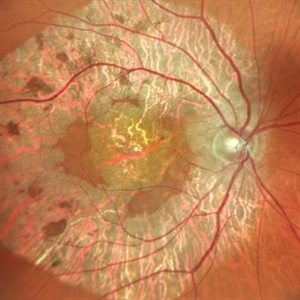
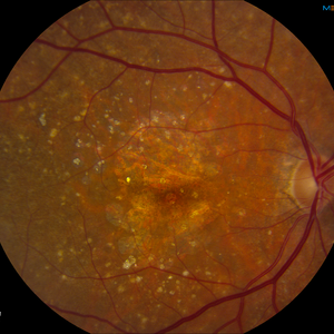 AMD with Calcific Drusen and Geographic Atrophy
Apr 19 2013 by Brandon G. Busbee, MD
AMD with calcific drusen and geographic atrophy.
Photographer: Alecia Camp, CRA - Tennessee Retina - Nashville, TN
Imaging device: Topcon TRC 50-EX
Condition/keywords: geographic atrophy
-
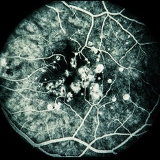
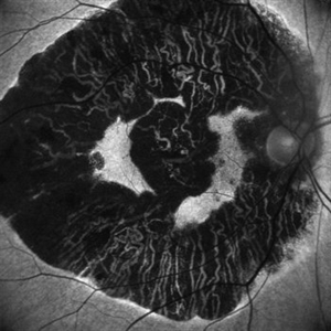
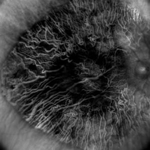
 Geographic Atrophy
Oct 13 2012 by Geoffrey G. Emerson, MD, PhD, FASRS
Geographic atrophy
Condition/keywords: advanced geographic atrophy, choroid, dry age-related macular degeneration (dry AMD)
-
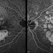
 Geographic Atrophy 2nd to Central areolar choroidal dystrophy
Nov 24 2012 by Roy Schwartz, MD
Fundus autofluorescence and SD-OCT of a 70-year-old woman with geographic atrophy sec. to Central areolar choroidal dystrophy
Condition/keywords: central areolar choroidal dystrophy (CACD), geographic atrophy
-
---thumb.jpg/image-square;max$300,300.ImageHandler) Age Related Macular Degeneration - Geographic Atrophy
Age Related Macular Degeneration - Geographic Atrophy
May 3 2013 by Suber S. Huang, MD, MBA, FASRS
Geographic Atrophy.
Imaging device: Retina Diseases Imaging Analysis Reading Center
Condition/keywords: advanced geographic atrophy, atrophic scar, atrophic spot, geographic atrophy, macula lesion, pigment epithelial atrophy
-
 Geographic Atrophy
Mar 27 2013 by Michael P. Kelly, FOPS
This is a combined FAF/SD-OCT in EDI mode of a patient with geographic atrophy and foveal sparing.
Photographer: Michael P. Kelly, FOPS. Director, Duke Eye Labs, Duke University Eye Center
Imaging device: Heidelberg Spectralis
Condition/keywords: enhanced depth imaging, foveal sparing, fundus autofluorescence (FAF), geographic atrophy, optical coherence tomography (OCT)
-
 Geographic Atrophy - Case 1: Photo 1 of 6
Oct 4 2012 by Gregg T. Kokame, MD, MMM, FASRS
NIRAF (near infrared autofluorescence) Image of patient with Geographic Atrophy
Photographer: Jaclyn Pisano, Retina Consultants of Hawaii
Imaging device: Heidelberg Spectralis
Condition/keywords: autofluorescence imaging, geographic atrophy, near infrared autofluorescence (NIRAF)
-
 Geographic Atrophy - Case 1: Photo 2 of 6
Oct 4 2012 by Gregg T. Kokame, MD, MMM, FASRS
Blue-peak Autofuorescence Image of patient with Geographic Atrophy
Photographer: Jaclyn Pisano, Retina Consultants of Hawaii
Imaging device: Heidelberg Spectralis
Condition/keywords: autofluorescence imaging, geographic atrophy
-
 Grouped Albinotic Spots in Macula
Mar 4 2014 by David Callanan, MD
39-year-old female, 20/15 OU; normal color, ERG, EOG, thresholds, VF.
Condition/keywords: albinism, macula
-
 Macular Degeneration with Extensive Geographic Atrophy
Jan 26 2022 by Olivia Rainey
Heidelberg Spectralis fluorescein angiography of a 94-year-old woman with Macular Degeneration affecting both eyes. The FA reveals transmission defects consistent with RPE changes and geographic atrophy of RPE of both eyes, as well as window defects consistent with peripheral scarring in the right eye. The patient's vision was Dcc20/70 in both eyes at the visit the images were taken.
Photographer: Olivia Rainey, OCT-C, COA
Imaging device: Heidelberg Spectralis
Condition/keywords: 30-degrees, choroidal neovascularization (CNV), dry age-related macular degeneration (dry AMD), early phase, fluorescein angiogram (FA), geographic atrophy, heidelberg spectralis, macular degeneration, neovascular age-related macular degeneration (AMD)
-
 Neovascular AMD with Active CNV
May 22 2025 by Kimberly Wakester
Optomap RGB of an 82-year-old man with Neovascular AMD with Active CNV and Dry AMD in the right eye. There is advanced atrophic changes without subfoveal involvement located temporally to the fovea. Patient is to continue follow up care with dilated exam, repeat OCT, and treatment of intravitreal injection of Vabysmo every 5 weeks at this time.
Photographer: Kimberly Wakester, COA, OCT-C
Imaging device: Optos California
Condition/keywords: advanced geographic atrophy, dry age-related macular degeneration (dry AMD), neovascular age-related macular degeneration (AMD)
-
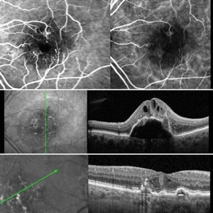 Retinal Angiomatous Proliferation RAP
Mar 11 2020 by RAFAEL REIS PEREIRA, MD
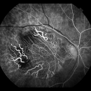
Retinal angiomatous proliferation (RAP) is a unique variant of neovascular age-related macular degeneration. Published studies have estimated that up to 15% of patients with neovascular age-related macular degeneration have RAP. Clinical features frequently associated with RAP include bilateral disease, presence of pigment epithelial detachments, and reticular pseudodrusen. RAP is more frequently associated with the development of retinal pigment epithelial tears and geographic atrophy that can lead to severe vision loss. We present a stereo fluorescein angiography and ICG (upper right and left image respectively) and OCT of left and right eye (middle and inferior image) of a RAP choroidal neovascularization in an 89-year-old patient.
Photographer: Rafael Reis Pereira
Imaging device: HRA Heildelberg Spectralis
Condition/keywords: retinal angiomatous proliferation (RAP)
-
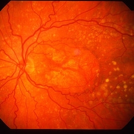
 Geographic Atrophy
Aug 29 2012 by Young Hee Yoon, MD, PhD
Fundus photograph of an 78-year-old woman. Her best-corrected visual acuity was counting fingers at 30cm.
Photographer: Kyoung Woon Kim, Asan Medical Center
Imaging device: Canon CR-DGI/Version 5.1.2
Condition/keywords: dry age-related macular degeneration (dry AMD), geographic atrophy
-
 Geographic Atrophy, Fundus photograph
Aug 23 2012 by Gerardo Garcia-Aguirre, MD
Fundus photograph of an 85-year-old patient with age related macular degeneration and geographic atrophy. A large area with well-defined borders is observed, in which the choroidal vasculature is visualized.
Photographer: Noemí Hernández, Asociación para Evitar la Ceguera en México
Imaging device: Zeiss FF4
Condition/keywords: geographic atrophy
-
---thumb.jpg/image-square;max$300,300.ImageHandler) Age Related Macular Degeneration - Geographic Atrophy
Age Related Macular Degeneration - Geographic Atrophy
May 3 2013 by Suber S. Huang, MD, MBA, FASRS
Geographic Atrophy.
Imaging device: Retina Diseases Imaging Analysis Reading Center
Condition/keywords: advanced geographic atrophy, atrophic scar, atrophic spot, geographic atrophy, macula lesion, pigment epithelial atrophy
-
 Dry Age-Related Macular Degeneration
Mar 29 2013 by Henry J. Kaplan, MD
Fundus photograph of a patient with dry AMD demonstrates multiple drusen, RPE change and geographic atrophy; notice that the patient has also familial or dominant drusen most prominant in nasal retina.
Condition/keywords: age-related macular degeneration (AMD), dry age-related macular degeneration (dry AMD), geographic atrophy
-
---thumb.jpg/image-square;max$300,300.ImageHandler) Geographic atrophy
Geographic atrophy
Aug 29 2012 by Young Hee Yoon, MD, PhD
OCT image of an 78-year-old woman. Her best-corrected visual acuity was counting fingers at 30cm.
Photographer: Ji Hee Kim, Asan Medical Center
Imaging device: Heidelberg spectralis
Condition/keywords: dry age-related macular degeneration (dry AMD), geographic atrophy
-
 Geographic Atrophy
May 11 2020 by Gayathri Mohan
Fundus autofluorescence image of a patient with geographic atrophy.
Photographer: Gayathri Mohan, Retina Foundation
Imaging device: Mirante, Nidek
Condition/keywords: fundus autofluorescence (FAF), geographic atrophy
-
 Geographic Atrophy
May 10 2020 by Gayathri Mohan
Color fundus photograph of a patient with geographic atrophy.
Photographer: Gayathri Mohan, Retina Foundation
Imaging device: Mirante, Nidek
Condition/keywords: fundus photograph, geographic atrophy
-
 Geographic Atrophy- Retro Mode
May 10 2020 by Gayathri Mohan
Image of a patient with geographic atrophy taken in retro mode. The deeper choroidal vessels are prominently seen.
Photographer: Gayathri Mohan, Retina Foundation
Imaging device: Mirante, Nidek
Condition/keywords: geographic atrophy, retro
-
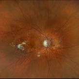
 GHPC
GHPC
May 13 2013 by Howard Schatz, MD
49-year-old white male, III GHPC, right eye: 10/200; left eye: 20/50.
Condition/keywords: geographic helicoid peripapillary choroidopathy (GHPC)
-
---thumb.jpg/image-square;max$300,300.ImageHandler) Abnormal Fundus
Abnormal Fundus
Oct 15 2013 by Maurice F. Rabb
The patient is a 48 year old female who was noted to have an abnormal fundus on routine examination. Her past medical history is unremarkable. Her past family history is remarkable for her father being diagnosed with macular degeneration at age 72. Visual acuity was 20/20-1, J-2, OD and 20/20, J-1, OS. Amsler grid examinaton was normal, OU. All 6 of the AOHRR screening plates were identified correctly, OU. The 45 minute rod psychophysiologic threshold was normal, OU. ERG responses were normal for both rod and cone amplitudes. The cone implicit times were normal. The EOG was normal.
Condition/keywords: abnormal fundus
 Loading…