Search results (307 results)
-
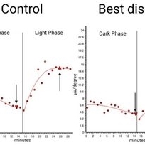 Representative Electrooculogram Responses
May 13 2024 by Gabrielle Hallai
Electrooculogram responses on the left from a control individual with no known retinal pathology. There is a clear dark trough around 13 minutes (arrow down) and a light peak around 25 minutes (arrow up). The Arden ratio, or the light peak to dark trough ratio, is 2.54, indicative of normal retinal pigment epithelium function (normal > 1.80, abnormal < 1.65). On the right-hand side, there is a representative image from an individual with Best macular dystrophy. Note the reduced responses for both the dark and light phase. There is a reduced Arden ratio of 1.23, suggestive of abnormal retinal pigment epithelium function. An abnormal Arden ratio is universal in Best vitelliform macular dystrophy and is the most common electroretinographic change in this disease. Other bestrophinopathies such as autosomal recessive bestrophinopathy may have normal EOG. EOG testing was completed on the Diagnosys ColorDome.
Photographer: Gabrielle Hallai, PhD, Cleveland Clinic Cole Eye Institute
Imaging device: Diagnosys ColorDome
Condition/keywords: Best disease, electrooculogram, electroretinography, EOG
-
---thumb.jpg/image-square;max$300,300.ImageHandler) Abnormal Fundus
Abnormal Fundus
Oct 15 2013 by Maurice F. Rabb
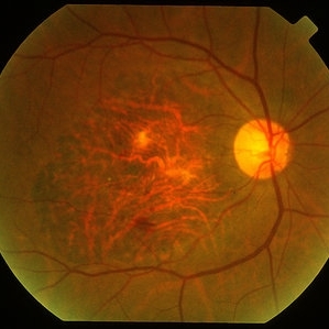
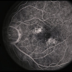
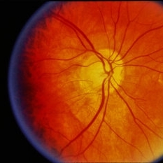
The patient is a 48 year old female who was noted to have an abnormal fundus on routine examination. Her past medical history is unremarkable. Her past family history is remarkable for her father being diagnosed with macular degeneration at age 72. Visual acuity was 20/20-1, J-2, OD and 20/20, J-1, OS. Amsler grid examinaton was normal, OU. All 6 of the AOHRR screening plates were identified correctly, OU. The 45 minute rod psychophysiologic threshold was normal, OU. ERG responses were normal for both rod and cone amplitudes. The cone implicit times were normal. The EOG was normal.
Condition/keywords: abnormal fundus
-
---thumb.jpg/image-square;max$300,300.ImageHandler) Abnormal Fundus
Abnormal Fundus
Oct 15 2013 by Maurice F. Rabb
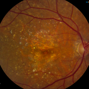
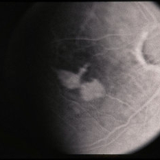
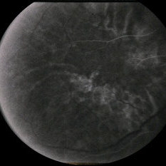
The patient is a 48 year old female who was noted to have an abnormal fundus on routine examination. Her past medical history is unremarkable. Her past family history is remarkable for her father being diagnosed with macular degeneration at age 72. Visual acuity was 20/20-1, J-2, OD and 20/20, J-1, OS. Amsler grid examinaton was normal, OU. All 6 of the AOHRR screening plates were identified correctly, OU. The 45 minute rod psychophysiologic threshold was normal, OU. ERG responses were normal for both rod and cone amplitudes. The cone implicit times were normal. The EOG was normal.
Condition/keywords: abnormal fundus
-
---thumb.jpg/image-square;max$300,300.ImageHandler) Abnormal Fundus
Abnormal Fundus
Oct 15 2013 by Maurice F. Rabb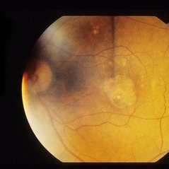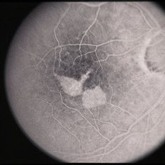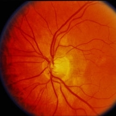
The patient is a 48 year old female who was noted to have an abnormal fundus on routine examination. Her past medical history is unremarkable. Her past family history is remarkable for her father being diagnosed with macular degeneration at age 72. Visual acuity was 20/20-1, J-2, OD and 20/20, J-1, OS. Amsler grid examinaton was normal, OU. All 6 of the AOHRR screening plates were identified correctly, OU. The 45 minute rod psychophysiologic threshold was normal, OU. ERG responses were normal for both rod and cone amplitudes. The cone implicit times were normal. The EOG was normal.
Condition/keywords: abnormal fundus
-
 Age Related Macular Degeneration
Mar 29 2013 by Henry J. Kaplan, MD
Geographic atrophy with small hemorrhages due to subretinal neovascular membrane development.
Condition/keywords: choroidal neovascularization (CNV), geographic atrophy
-
---thumb.jpg/image-square;max$300,300.ImageHandler) Age Related Macular Degeneration
Age Related Macular Degeneration
May 3 2013 by Suber S. Huang, MD, MBA, FASRS
Age related macular degeneration.
Condition/keywords: advanced geographic atrophy, atrophic scar, atrophic spot, geographic atrophy, macula lesion, pigment epithelial atrophy, red-free, window defect
-
---thumb.jpg/image-square;max$300,300.ImageHandler) Age Related Macular Degeneration - Geographic Atrophy
Age Related Macular Degeneration - Geographic Atrophy
May 3 2013 by Suber S. Huang, MD, MBA, FASRS
Geographic Atrophy.
Imaging device: Retina Diseases Imaging Analysis Reading Center
Condition/keywords: advanced geographic atrophy, atrophic scar, atrophic spot, geographic atrophy, macula lesion, pigment epithelial atrophy
-
---thumb.jpg/image-square;max$300,300.ImageHandler) Age Related Macular Degeneration - Geographic Atrophy
Age Related Macular Degeneration - Geographic Atrophy
May 3 2013 by Suber S. Huang, MD, MBA, FASRS
Geographic Atrophy.
Imaging device: Retina Diseases Imaging Reading Center
Condition/keywords: advanced geographic atrophy, atrophic scar, atrophic spot, geographic atrophy, macula lesion, pigment epithelial atrophy, red-free, window defect
-
---thumb.jpg/image-square;max$300,300.ImageHandler) Age Related Macular Degeneration - Geographic Atrophy
Age Related Macular Degeneration - Geographic Atrophy
May 3 2013 by Suber S. Huang, MD, MBA, FASRS
Geographic Atrophy.
Imaging device: Retina Diseases Imaging Analysis Reading Center
Condition/keywords: advanced geographic atrophy, atrophic scar, atrophic spot, geographic atrophy, macula lesion, pigment epithelial atrophy
-
 AMD - Geographic Atrophy
Oct 12 2012 by Gregg T. Kokame, MD, MMM, FASRS
AMD, geographic atrophy
Photographer: Jaclyn Pisano, Retina Consultants of Hawaii
Imaging device: Zeiss FF-450 plus
Condition/keywords: geographic atrophy
-
 AMD with Calcific Drusen and Geographic Atrophy
Apr 19 2013 by Brandon G. Busbee, MD
AMD with calcific drusen and geographic atrophy.
Photographer: Alecia Camp, CRA - Tennessee Retina - Nashville, TN
Imaging device: Topcon TRC 50-EX
Condition/keywords: geographic atrophy
-
 AMPPPE
Apr 16 2013 by Howard Schatz, MD
III AMPPPE and GHPC; right eye: 20/20; left eye: 20/80.
Condition/keywords: acute posterior multifocal placoid pigment epitheliopathy (APMPPE), geographic helicoid peripapillary choroidopathy (GHPC)
-
 ARMD
ARMD
Aug 7 2013 by H. Michael Lambert, MD
ARMD. Geographic atrophy
-
 ARMD - Geographic Atrophy
Feb 26 2014 by David Callanan, MD
Age-related macular degeneration (AMD) geographic atrophy.
Condition/keywords: age-related macular degeneration (AMD), geographic atrophy
-
 ARMD - Geographic Atrophy
Feb 26 2014 by David Callanan, MD
Age-related macular degeneration (AMD) geographic atrophy.
Condition/keywords: age-related macular degeneration (AMD), geographic atrophy
-
 ARMD - Geographic Atrophy
Feb 26 2014 by David Callanan, MD
Age-related macular degeneration (AMD) geographic atrophy.
Condition/keywords: age-related macular degeneration (AMD), geographic atrophy
-
 ARMD - Geographic Atrophy
Feb 26 2014 by David Callanan, MD
Age-related macular degeneration (AMD) geographic atrophy.
Condition/keywords: age-related macular degeneration (AMD), geographic atrophy
-
 ARMD - Geographic Atrophy
Feb 26 2014 by David Callanan, MD
Age-related macular degeneration (AMD) geographic atrophy.
Condition/keywords: age-related macular degeneration (AMD), geographic atrophy
-
 ARMD - Geographic Atrophy
Feb 26 2014 by David Callanan, MD
Age-related macular degeneration (AMD) geographic atrophy.
Condition/keywords: age-related macular degeneration (AMD), geographic atrophy
-
 ARMD - Geographic Atrophy
Feb 26 2014 by David Callanan, MD
Age-related macular degeneration (AMD) geographic atrophy.
Condition/keywords: age-related macular degeneration (AMD), geographic atrophy
 Loading…