Search results (307 results)
-
 White with Pressure Phenomenon
Nov 9 2012 by Norman Byer
This photograph shows a rather typical example of the white with pressure phenomenon. It may take many forms but usually has a geographic configuration with irregular borders which may be rounded or angular. Its cause is unknown but it is a very common finding in the fundus. It is important to know that it does not indicate the presence of any disease of the retina. Over a period of time, it may disappear completely or it may change its configuration or location. Other examples of this phenomenon are shown in slide pairs 81 and 103.
Condition/keywords: geographic configuration, irregular borders, white retinal lesion, white with pressure
-
 Geographic Atrophy, Fundus photograph
Aug 23 2012 by Gerardo Garcia-Aguirre, MD
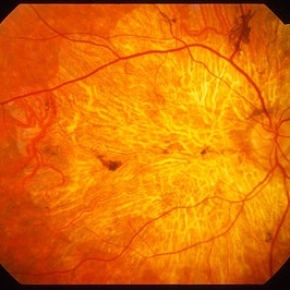
Fundus photograph of an 85-year-old patient with age related macular degeneration and geographic atrophy. A large area with well-defined borders is observed, in which the choroidal vasculature is visualized.
Photographer: Noemí Hernández, Asociación para Evitar la Ceguera en México
Imaging device: Zeiss FF4
Condition/keywords: geographic atrophy
-
---thumb.jpg/image-square;max$300,300.ImageHandler) Geographic atrophy
Geographic atrophy
Aug 29 2012 by Young Hee Yoon, MD, PhD
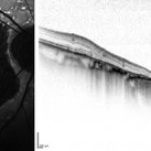
OCT image of an 78-year-old woman. Her best-corrected visual acuity was counting fingers at 30cm.
Photographer: Ji Hee Kim, Asan Medical Center
Imaging device: Heidelberg spectralis
Condition/keywords: dry age-related macular degeneration (dry AMD), geographic atrophy
-
 Geographic Atrophy
Aug 29 2012 by Young Hee Yoon, MD, PhD
Fundus photograph of an 78-year-old woman. Her best-corrected visual acuity was counting fingers at 30cm.
Photographer: Kyoung Woon Kim, Asan Medical Center
Imaging device: Canon CR-DGI/Version 5.1.2
Condition/keywords: dry age-related macular degeneration (dry AMD), geographic atrophy
-
 AMD with Calcific Drusen and Geographic Atrophy
Apr 19 2013 by Brandon G. Busbee, MD
AMD with calcific drusen and geographic atrophy.
Photographer: Alecia Camp, CRA - Tennessee Retina - Nashville, TN
Imaging device: Topcon TRC 50-EX
Condition/keywords: geographic atrophy
-
 ARMD With Geographic Atrophy, Peripheral Degeneration
Dec 6 2013 by James B. Soque, CRA, OCT-C, COA, FOPS
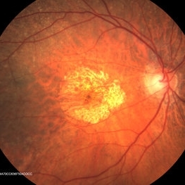
92-year-old white female with exudative macular degeneration, geographic atrophy, and peripheral retinal degeneration.
Photographer: James Soque, CRA COA, Island Retina, Shirley, New York
Imaging device: Topcon TRC 50DX with OIS 10.6.45
Condition/keywords: geographic atrophy, macular degeneration, retinal degeneration
-
---thumb.jpg/image-square;max$300,300.ImageHandler) Age Related Macular Degeneration - Geographic Atrophy
Age Related Macular Degeneration - Geographic Atrophy
May 3 2013 by Suber S. Huang, MD, MBA, FASRS
Geographic Atrophy.
Imaging device: Retina Diseases Imaging Analysis Reading Center
Condition/keywords: advanced geographic atrophy, atrophic scar, atrophic spot, geographic atrophy, macula lesion, pigment epithelial atrophy
-
---thumb.jpg/image-square;max$300,300.ImageHandler) Primary Hyperoxaluria and Oxalosis
Primary Hyperoxaluria and Oxalosis
Jul 24 2013 by Hamid Ahmadieh, MD
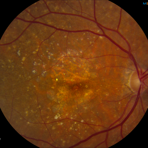
Color fundus photograph of the right eye of a 55-year-old man with primary hyperoxaluria and oxalosis. Characteristic crystalline retinopathy (flecked retina), black geographic maculopathy, and partial optic atrophy are visible. In addition, occluded branches of central retinal artery due to calcium oxalate deposition are visible.
Photographer: Hanieh Payab, Ophthalmic Research Center, Labbafinejad Medical Center, Tehran
Imaging device: Topcon Fundus Camera
Condition/keywords: oxalosis, primary hyperoxaluria
-
 Central Areolar Choroidal Dystrophy
Apr 14 2013 by Edwin H. Ryan, MD
Fundus photograph of a 52-year-old woman with CACD. 5/200 OD, 20/50 OS.
Condition/keywords: central areolar choroidal dystrophy (CACD), geographic atrophy, macula
-
 Berlin's Edema
Apr 8 2019 by Gary R. Cook, MD, FACS
39-year-old white female with geographic area of retinal whitening ( Berlin's edema) without hemorrhage in the midperiphery secondary to blunt trauma; V.A. = 20/25
Imaging device: Topcon VT-50
Condition/keywords: Berlin's edema, blunt trauma, retinal edema
-
---thumb.jpg/image-square;max$300,300.ImageHandler) Age Related Macular Degeneration - Geographic Atrophy
Age Related Macular Degeneration - Geographic Atrophy
May 3 2013 by Suber S. Huang, MD, MBA, FASRS
Geographic Atrophy.
Imaging device: Retina Diseases Imaging Analysis Reading Center
Condition/keywords: advanced geographic atrophy, atrophic scar, atrophic spot, geographic atrophy, macula lesion, pigment epithelial atrophy
-
 Geographic Atrophy - Case 1: Photo 2 of 6
Oct 4 2012 by Gregg T. Kokame, MD, MMM, FASRS
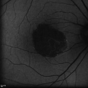
Blue-peak Autofuorescence Image of patient with Geographic Atrophy
Photographer: Jaclyn Pisano, Retina Consultants of Hawaii
Imaging device: Heidelberg Spectralis
Condition/keywords: autofluorescence imaging, geographic atrophy
-
 Geographic atrophy
Aug 29 2012 by Young Hee Yoon, MD, PhD
FAF image of an 78-year-old woman. Her best-corrected visual acuity was counting fingers at 30cm.
Photographer: Kyoung Woon Kim, Asan Medical Center
Imaging device: Heidelberg
Condition/keywords: dry age-related macular degeneration (dry AMD), geographic atrophy
-
 Central Areolar Choroidal Dystrophy
Apr 14 2013 by Edwin H. Ryan, MD
Fundus photograph of a 52-year-old woman with CACD. 5/200 OD, 20/50 OS.
Condition/keywords: central areolar choroidal dystrophy (CACD), geographic atrophy, macula
-
---thumb.jpg/image-square;max$300,300.ImageHandler) Geographic Atrophy - Fundus Photograph
Geographic Atrophy - Fundus Photograph
Oct 3 2013 by Gerardo Garcia-Aguirre, MD
Geographic atrophy - fundus photograph.
Condition/keywords: fundus photograph, geographic atrophy
-
 Geographic Atrophy
Oct 13 2012 by Geoffrey G. Emerson, MD, PhD, FASRS
Geographic atrophy
Condition/keywords: advanced geographic atrophy, choroid, dry age-related macular degeneration (dry AMD)
-
 Geographic Atrophy - Case 1: Photo 3 of 6
Oct 4 2012 by Gregg T. Kokame, MD, MMM, FASRS
Infrared Image of patient with Geographic Atrophy
Photographer: Jaclyn Pisano, Retina Consultants of Hawaii
Imaging device: Heidelberg Spectralis
Condition/keywords: autofluorescence imaging, geographic atrophy
-
 Geographic Atrophy 2nd to Central areolar choroidal dystrophy
Nov 24 2012 by Roy Schwartz, MD
Fundus autofluorescence and SD-OCT of a 70-year-old woman with geographic atrophy sec. to Central areolar choroidal dystrophy
Condition/keywords: central areolar choroidal dystrophy (CACD), geographic atrophy
-
 Geographic Atrophy - Case 1: Photo 4 of 6
Oct 4 2012 by Gregg T. Kokame, MD, MMM, FASRS
NIRAF (near infrared autofluorescence) Image of patient with Geographic Atrophy
Photographer: Jaclyn Pisano, Retina Consultants of Hawaii
Imaging device: Heidelberg Spectralis
Condition/keywords: autofluorescence imaging, geographic atrophy, near infrared autofluorescence (NIRAF)
-
 cRORA
cRORA
Aug 5 2020 by Dhaivat Shah
A 54-year-old healthy male presented to us with a decreased vision in right eye since past 8 years. The patient gave a history of bleed in right eye before 8 years for which some intravitreal injection was given; post which there no major visual improvement. No details or documentation was available regarding the same. His BCVA in the right eye was 5/60. Fundus examination revealed a sharply demarcated hypopigmented patch over the macula with mild posterior excavation suggestive of macular scar. OCT image shows foveal thinning with loss of Retinal pigment epithelium and outer retinal layers (RORA). There are 2 types of RORAs, complete and incomplete. Complete RORA and incomplete RORA are entities defined by various imaging modalities describing atrophy of the retinal pigment epithelial and the outer retinal layers. OCT imaging defines incomplete RORA (iRORA) as a region of signal hyper transmission into the choroid and a corresponding zone of attenuation ordisruption of the RPE (<250um) and evidence of overlying photoreceptor degeneration (<250um). There should not be any RPE tear associated with it. OCT imaging describes complete RORA (cRORA) based on 4 inclusion criteria. These include, area of hypertransmission of more than 250um, zone of attenuation or disruption of the RPE of more than 250um in diameter, evidence of overlying photoreceptor degeneration and absence of scrolled RPE or other signs of an RPE tear. Other modalities used to define these include fundus autoflourescence(FAF), near infrared reflectance(NIR) and color fundus photograph(CFP). On CFP, it shows a sharply demarcated hypopigmented of >250um size with better visibility of choroidal vessels. FAF shows a hypo autoflourescent patch with sharply demarcated borders of size >250um, the colour of which is similar to that of the optic nerve head or retinal blood vessels excluding any pigmentation or artefact. On NIR, it shows a hyperreflective area with sharply demarcated borders of >250um size excluding any artefact. RORA can be seen in conditions like geographical atrophy in ARMD, central areolar choroidal dystrophy, atrophy secondary to anti-VEGF treatment. References: 1. Sadda SR, Guymer R, Holz FG, et al. Consensus Definition for Atrophy Associated with Age-Related Macular Degeneration on OCT: Classification of Atrophy Report 3 [published correction appears in Ophthalmology. 2019 Jan;126(1):177]. Ophthalmology. 2018;125(4):537-548. 2. Guymer RH, Rosenfeld PJ, Curcio CA, et al. Incomplete Retinal Pigment Epithelial and Outer Retinal Atrophy in Age-Related Macular Degeneration: Classification of Atrophy Meeting Report 4. Ophthalmology. 2020;127(3):394-409. 3. Eng VA, Rayess N, Nguyen HV, Leng T. Complete RPE and outer retinal atrophy in patients receiving anti-VEGF treatment for neovascular age-related macular degeneration. PLoS One. 2020;15(5):e0232353.
Photographer: Miss Anjum Zafar Khan
Imaging device: Choithram Netralaya
Condition/keywords: macular scar, outer retina, retinal pigment epithelium
-
 Geographic Atrophy
Mar 29 2013 by Henry J. Kaplan, MD
Large GA involving the whole posterior pole.
Condition/keywords: dry age-related macular degeneration (dry AMD), geographic atrophy
-
 Geographic Atrophy - Colour Image
Sep 27 2013 by Gerardo Garcia-Aguirre, MD
Geographic atrophy.
Condition/keywords: color photo, geographic atrophy
-
 Geographic Atrophy - Case 1: Photo 1 of 6
Oct 4 2012 by Gregg T. Kokame, MD, MMM, FASRS
NIRAF (near infrared autofluorescence) Image of patient with Geographic Atrophy
Photographer: Jaclyn Pisano, Retina Consultants of Hawaii
Imaging device: Heidelberg Spectralis
Condition/keywords: autofluorescence imaging, geographic atrophy, near infrared autofluorescence (NIRAF)
-
---thumb.jpg/image-square;max$300,300.ImageHandler) Abnormal Fundus
Abnormal Fundus
Oct 15 2013 by Maurice F. Rabb
The patient is a 48 year old female who was noted to have an abnormal fundus on routine examination. Her past medical history is unremarkable. Her past family history is remarkable for her father being diagnosed with macular degeneration at age 72. Visual acuity was 20/20-1, J-2, OD and 20/20, J-1, OS. Amsler grid examinaton was normal, OU. All 6 of the AOHRR screening plates were identified correctly, OU. The 45 minute rod psychophysiologic threshold was normal, OU. ERG responses were normal for both rod and cone amplitudes. The cone implicit times were normal. The EOG was normal.
Condition/keywords: abnormal fundus
-
 ARMD With Geographic Atrophy , Peripheral Degeneration, FA
Dec 6 2013 by James B. Soque, CRA, OCT-C, COA, FOPS
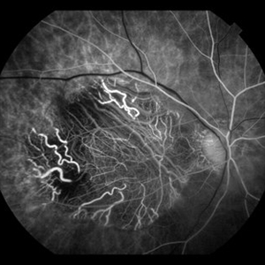
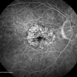
FA right eye early phase, of a 92-year-old white female with exudative macular degeneration, geographic atrophy, and peripheral retinal degeneration.
Photographer: James Soque CRA COA, Island Retina, Shirley, New York
Imaging device: Topcon TRC 50DX with OIS 10.6.45
Condition/keywords: geographic atrophy
 Loading…