Search results (1240 results)
-
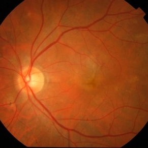
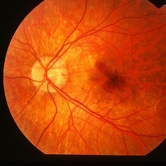
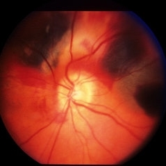
 Optic Nerve Head Drusen With Idiopathic CNV
Feb 17 2017 by Kristen Wagner
22-year-old female fundus photograph of a right eye with Optic Nerve Drusen with Idiopathic CNV.
Photographer: Kristen Wagner, COT, OSC Ophthalmic Photographer, Tennessee Retina, Nashville TN
Condition/keywords: choroidal neovascularization (CNV), drusen of optic disc, optic disc drusen
-
 Active CNVM
Jul 11 2016 by Manish Nagpal, MD, FRCS (UK), FASRS
Colour photo showing an active CNVM.
Photographer: pooja barot
Condition/keywords: choroidal neovascular membrane (CNVM), optical coherence tomography (OCT)
-
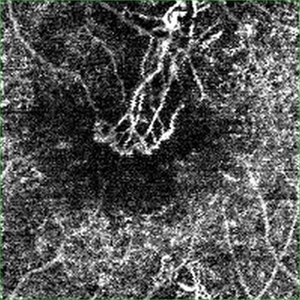 Active CNVM on Angio OCT
Jul 11 2016 by Manish Nagpal, MD, FRCS (UK), FASRS
Angio OCT picture showing neovascularization corresponding to the area of CNVM.
Photographer: pooja barot
Condition/keywords: choroidal neovascular membrane (CNVM), optical coherence tomography (OCT)
-
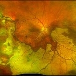
 Cat Eye Syndrome
Feb 11 2020 by Sophia El Hamichi, MD
A 3-year-old female with cat eye syndrome including iris, chorioretinal and optic nerve colobomas. Note the CNV temporally to the optic nerve coloboma (blue arrows)
Photographer: Giselle De Oliveira, Bascom Palmer Eye Institute, Miami
Imaging device: RetCam
Condition/keywords: cat eye syndrome, chorioretinal coloboma, choroidal neovascularization (CNV), coloboma, coloboma of optic disc, optic nerve coloboma
-
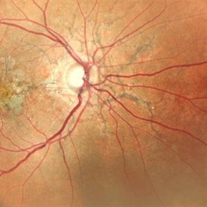
 Choroidal Neovascularization
May 27 2020 by Jamin S. Brown, MD
73-year-old female with CNV.
Photographer: Jeffrey Barker, Retina-Vitreous Surgeons of CNY
Condition/keywords: choroidal neovascularization (CNV)
-
 Peripheral CNVM with Extensive Scarring
Oct 12 2019 by John S. King, MD
82-year-old white male with an acute loss of vision in the right eye was sent in to rule out a retinal detachment. Vision was 20/350; a dense VH was present, b-scan showed irregular areas of high reflectivity in the periphery that was c/w SRH. Peripherally, a few weeks later, there were areas that could be seen and were c/w peripheral CNVM (old and new). Anti-VEGF was administered. A month later vision was unchanged and patient wanted surgery to remove the VH. Pictured is one week since surgery; large peripheral scars are seen; diffuse areas of SR pigmentation is present; vitreous skirt present; and a few IRHs secondary to DR can be seen. He is currently 20/70 sc.
Photographer: Shelly Blair
Imaging device: Optos CA
Condition/keywords: choroidal neovascular membrane (CNVM), peripheral fundus lesion, vitreous blood
-
---thumb.jpg/image-square;max$300,300.ImageHandler) Active Choroidal Neovascularization With Subretinal Hemorrhage
Active Choroidal Neovascularization With Subretinal Hemorrhage
Nov 25 2013 by Maurice F. Rabb
Active choroidal neovascularization with subretinal hemorrhage.
Condition/keywords: choroidal neovascularization (CNV), subretinal hemorrhage
-
 Angioid Streaks
Jan 20 2021 by Nivesh Gupta
Fundus photograph of an 51-year-old female patient with angioid streaks with secondary choroidal neovascular membrane.
Photographer: Nivesh Gupta, Retina Fellow, Retina Foundation, Ahmedabad, India
Imaging device: NIDEK SLO MIRANTE
Condition/keywords: age-related macular degeneration (AMD), angioid streaks, choroidal neovascular membrane (CNVM)
-
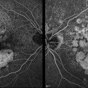
 Central Serous Chorioretinopathy
Jan 25 2022 by Olivia Rainey
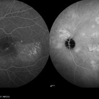
Late phase widefield fluorescein angiography of a 60-year-old male with Central Serous Chorioretinopathy. Chronic history of CSR followed with observation without treatment prior to presenting at our office. The physician noted subfoveal subretinal fluid with recent visual decline. FA shows multifocal leakage and ICG shows hypercyanescence. OCTA, ICG, and FA consistent with CSR, and without concern for CNVM thus will observe without anti-VEGF at this time. PDT therapy recommended.
Photographer: Olivia Rainey, OCT-C, COA
Imaging device: Heidelberg Spectralis
Condition/keywords: 55-degrees, central serous chorioretinopathy (CSCR), central serous retinopathy (CSR), chronic central serous chorioretinopathy (CSCR), fluorescein angiogram (FA), heidelberg spectralis, indocyanine green (ICG) angiography, left eye
-
 Choroidal Osteoma Plus CNV
Sep 2 2012 by Hamid Ahmadieh, MD
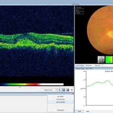
Color fundus photograph and OCT imaging of a 47-year-old man with a juxtafoveal CNV superimposed on a choroidal osteoma.
Photographer: Hamid Ahmadieh, Ophthalmic Research Center, Labbafinejad Medical Center
Imaging device: Topcon
Condition/keywords: choroidal neovascularization (CNV), choroidal osteoma, optical coherence tomography (OCT)
-
 CNV due to AMPPE
Oct 16 2012 by Ratimir Lazic, MD, PhD
FAG of 58-year-old male. In early venous phase hyperflorescence of white dots (caused by window defect) can be seen. Leakage of dye in juxtafoveolar region.
Photographer: Marko Lukic, MD
Imaging device: Zeis Visucam Lite 2
Condition/keywords: acute posterior multifocal placoid pigment epitheliopathy (APMPPE), choroidal neovascularization (CNV)
-
 CNV due to AMPPE
Oct 16 2012 by Ratimir Lazic, MD, PhD
Color fundus photography of a 58-year-old male. White dots with juxtafoveolar subretinal fluid can be seen. BCVA of that eye is 0.35.
Photographer: Marko Lukic, MD
Imaging device: Zeis Visucam Lite 2
Condition/keywords: acute posterior multifocal placoid pigment epitheliopathy (APMPPE), choroidal neovascularization (CNV)
-
 CNVM in Pan-retinal Photocoagulated Patient
Dec 30 2020 by ASRS Staff
Wide fundus photograph of 65-year-old, female, diabetic patient.
Imaging device: Nidek Mirante
Condition/keywords: age-related macular degeneration (AMD), diabetes, pan-retinal photocoagulation (PRP)
-
 Ehlers-Danlos Syndrome
Apr 22 2021 by Harita Shah
Fundus photograph of a 37-year-old male, known case of Ehlers-Danlos Syndrome, having left eye CNVM scar with angioid streaks.
Photographer: Harita Shah, Banker's Retina Clinic & Laser Centre
Imaging device: Topcon TRC 50DX
Condition/keywords: Ehlers-Danlos syndrome
-
 Idiopathic Choroidal Neovascularization
Mar 2 2023 by Corey Grant
Optical coherence tomography and ultra-wide field fundus photograph of a 51 year old male with idiopathic choroidal neovascularization affecting his right eye. The patient had no symptoms at the time of the appointment and his vision was Dcc20/20-2 OU. The physcian stated that there wasn't active exudation on the exam or ocular imaging and based on the clinical findings, he has recommended we defer any treatments.
Photographer: Corey Grant
Imaging device: Heidelberg Spectralis, OPTOS California
Condition/keywords: choroidal neovascularization (CNV), CNVM, fundus photograph, OCT, optical coherence tomography (OCT), Optos, Right Eye, ultra-wide field imaging
-
 Idiopathic Peripapillary CNV
Jan 4 2024 by Virginia Gebhart
13 year old female with inactive CNV. Increased pigment 360 at 1 year follow up. No inflammation or SRF, pt remains asymptomatic
Photographer: Virginia Gebhart
Imaging device: Optos California
Condition/keywords: choroidal neovascularization (CNV), peripapillary choroidal neovascularization (PPCNVM)
-
 Juxtafoveal Choroidal Neovascularization Secondary to Choroidal Rupture
Aug 30 2012 by Young Hee Yoon, MD, PhD
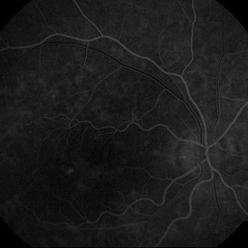
Fluorescence Angiography (FA) image of a 14-year-old boy with a history of blunt trauma to his left eye 9 months ago. Best-corrected visual acuity remained at 20/30.
Photographer: Heon Eui Hong, Asan Medical Center
Imaging device: HHeidelberg HRA II/ version 1.7.0.0
Condition/keywords: choroidal rupture, juxtafoveal choroidal neovascularization (CNV)
-
 Macular Degeneration with Extensive Geographic Atrophy
Jan 26 2022 by Olivia Rainey
Heidelberg Spectralis fluorescein angiography of a 94-year-old woman with Macular Degeneration affecting both eyes. The FA reveals transmission defects consistent with RPE changes and geographic atrophy of RPE of both eyes, as well as window defects consistent with peripheral scarring in the right eye. The patient's vision was Dcc20/70 in both eyes at the visit the images were taken.
Photographer: Olivia Rainey, OCT-C, COA
Imaging device: Heidelberg Spectralis
Condition/keywords: 30-degrees, choroidal neovascularization (CNV), dry age-related macular degeneration (dry AMD), early phase, fluorescein angiogram (FA), geographic atrophy, heidelberg spectralis, macular degeneration, neovascular age-related macular degeneration (AMD)
-
 Myopic CNV
May 2 2013 by Henry J. Kaplan, MD
Choroidal neovascularization with hemorrhage in a highly myopic patient.
Condition/keywords: myopic choroidal neovascularization (CNV)
-
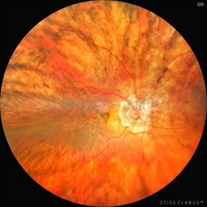 Myopic CNVM
Jul 22 2022 by T. P . VIGNESH, MBBS,MS
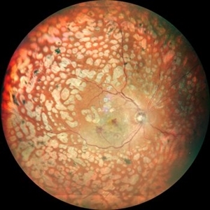
Fundus photograph of a 64-year-old woman with high myopia , myopic fundus and myopic CNVM.
Photographer: Bharathi Singaravel
Imaging device: Zeiss Clarus
Condition/keywords: high myopia, Myopic CNVM
-
 Neovascular AMD with Active CNV
May 22 2025 by Kimberly Wakester
Optomap RGB of an 82-year-old man with Neovascular AMD with Active CNV and Dry AMD in the right eye. There is advanced atrophic changes without subfoveal involvement located temporally to the fovea. Patient is to continue follow up care with dilated exam, repeat OCT, and treatment of intravitreal injection of Vabysmo every 5 weeks at this time.
Photographer: Kimberly Wakester, COA, OCT-C
Imaging device: Optos California
Condition/keywords: advanced geographic atrophy, dry age-related macular degeneration (dry AMD), neovascular age-related macular degeneration (AMD)
-
 Optic Nerve Pit OD - OCT
Aug 6 2018 by Hosam Attia, MD
65-year-old white male, presented for a second opinion for possible cataract extraction OD. BCVA: OD: 20/70 OS: 20/60 WRx: OD: -3.75 +1.50 x 5 OS: -1.75 +1.50 x 178 SLE: +2 NS OD>OS DFE: OD: Nasal macular GA, connected by milder track of RPE changes to an optic nerve pit OD (no fluid seen clinically) OS: enlarged C/D w/ no pits, macular RPE change w/ No heme, CME/ SRF OCT: OD: Peri-papillary cystoid changes & outer retinal atrophy (corresponding to the area of GA on the pseudocolor photo) w/ No SRF (mimicking PP CNVM), connected to the optic disc pit by shallow sinus/ tract. OS: Drusenoid RPE changes, No cystoid changes/ SRF
Imaging device: Zeiss Cirrus -5000
Condition/keywords: congenital optic nerve pit
-
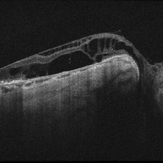
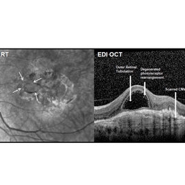 Outer Retinal Tubulation
Mar 27 2018 by Dhaivat Shah
Outer retinal tubulation (ORT) is a feature of photoreceptor rearrangement after chronic retinal damage due to refractory cme, long standing CNVM or old trauma. Photoreceptors lose adhesions to surrounding structures, resulting in outward folding and formation of new lateral contact between photoreceptors to form round structure. They generally remains stable over time. It is important to recognize ORT on OCT because it indicates a refractory state of the pathological condition and poor visual prognosis, and likely not to benefit from any treatment. Here is a case of 62-year-old female with history of 4 previous anti-VEGF injection in left eye for CNVM, with the recent OCT showing formation of ORT with subfoveal scarred membrane.
Photographer: Dr Dhaivat Shah
Condition/keywords: choroidal neovascular membrane (CNVM), outer retinal tubulation
-
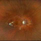
 Peripapillary CNVM - ARMD
Jan 16 2014 by David Callanan, MD
Peripapillary CNVM - ARMD, 81-old-year female.
Condition/keywords: peripapillary
-
 Pigment Epithelial Detachment late FA with small occult CNV
Jul 6 2012 by Tarek S. Hassan, MD, FASRS
72-year-old man with VA loss and metamorphopsia of 2 months duration. PED found, testing done to rule out CNV. Very suspicious for CNV in superonasal fovea/parafovea.
Condition/keywords: choroidal neovascularization (CNV), pigment epithelial detachment (PED)
 Loading…