 Initializing download.
-
 By Mateus Queiroz Corrêa, MD
Sorocaba Eye Bank Hospital - Uploaded on Jan 6, 2025.
- Last modified by Joshua Friedman on Jan 6, 2025.
- Rating
- Appears in
- Pneumatic Retinopexy
- Condition/keywords
- pneumatic retinopexy
- Photographer
- Mateus Queiroz Corrêa, Sorocada Eye Bank Hospital
- Imaging device
-
Fundus camera
Optos California - Description
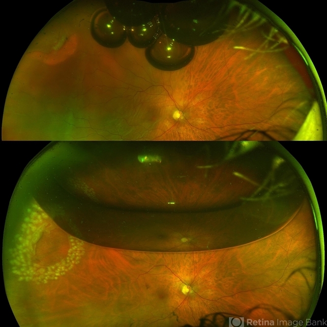
- Fundus photograph of a pneumatic retinopexy. The upper photo taken just 30 minutes after C3F8 gas injection shows rhegmatogenous retinal detachment with superior temporal horseshoe tear and gas bubbles resembling fish-eggs. After two days with appropriated head position (botton photo), the retina is attached and laser photocoagulation was performed on the border of the break. A Single great gas bubble was formed.


---thumb.JPG/image-square;max$79,0.ImageHandler "Gas Bubble")
---thumb.JPG/image-square;max$79,0.ImageHandler "Gas Bubble")


