 Initializing download.
-
 By Virginia Gebhart
Retina Consultants of Carolina
Co-author(s): Chris Bergstrom, MD, OD - Uploaded on Jul 3, 2025.
- Last modified by Virginia Gebhart on Aug 15, 2025.
- Rating
- Appears in
- Miscellaneous
- Condition/keywords
- lymphoma, biopsy, gas bubble
- Photographer
- Virginia Gebhart, Retina Consultants of Carolina
- Imaging device
-
Fundus camera
Optos California - Description
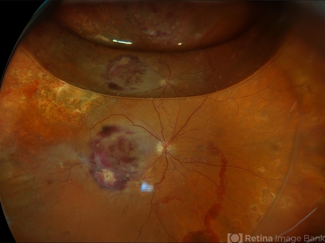
- 78 year old male s/p vitreous biopsy for T-Cell lymphoma. Pt presented with peripheral blot hemorrhages and numerous white subretinal infiltrates. Retinal pallor and thickening temporally. History of cutaneous T-cell lymphoma. PPV/vitreous biopsy performed to find differential diagnosis. Silicone oil was placed for 6 weeks, then removed and exchanged with a gas bubble. Hematology pathologist and Emory reviewed path report and agrees it is consistent with T-cell lymphoma. Pt received intravitreal Methotrexate and will be scheduled for weekly treatments. BCVA CF


---thumb.JPG/image-square;max$79,0.ImageHandler "Choroidal lymphoma in remission following radiotherapy")
---thumb.JPG/image-square;max$79,0.ImageHandler "Choroidal Lymphoma")

---thumb.JPG/image-square;max$79,0.ImageHandler "choroidal lymphoma")
---thumb.JPG/image-square;max$79,0.ImageHandler "choroidal lymphoma")
---thumb.JPG/image-square;max$79,0.ImageHandler "choroidal lymphoma")
")
---thumb.jpg/image-square;max$79,0.ImageHandler "Lymphoma Acute Retinal Necrosis (ARN)")
