Search results (3015 results)
-
 Unexpected Sanctuary: Gas Bubble Entrapment in Morning Glory Disc
Sep 5 2025 by Danny Salgado Gómez
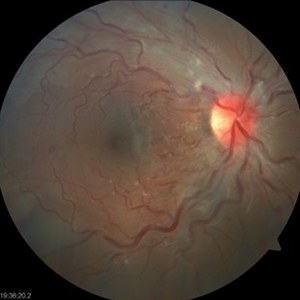
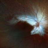
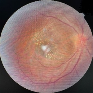
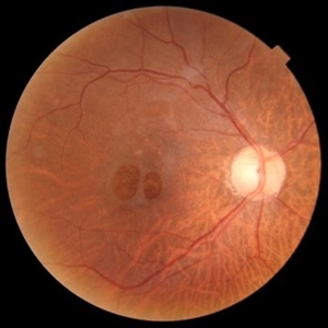
Fundus photograph of a 62-year-old male patient with Morning Glory syndrome in the right eye, who underwent vitrectomy, gas, and endolaser for posterior pole detachment. In the postoperative period, a gas bubble is observed within the optic disc, which persisted even after complete reabsorption of the intraocular gas.
Photographer: Dr. Danny Salgado, Retina and Vitreous Fellow, Clínica Oftalmológica del Caribe, Colombia.
Condition/keywords: gas bubble, intraocular gas, Morning Glory, Retinal Detachment, vitrectomy
-
 Papillophlebitis Salauno
Sep 3 2025 by Pablo Angel Garcia Uribe
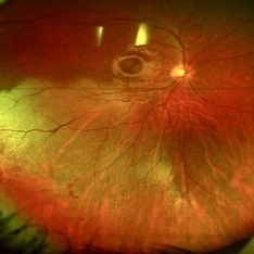
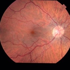
Fundus photograph of a 24-year-old woman, previously healthy, with a history of recreational inhaled cannabis use, presented with a 24-hour history of photopsias and mild decrease in visual acuity, associated with a subtle relative central scotoma in the right eye. On ophthalmic examination, the anterior segment of both eyes was unremarkable. Best-corrected visual acuity was slightly reduced in the right eye and normal in the left. Fundus biomicroscopy of the right eye revealed moderate disc edema with hyperemia and well-defined margins, accompanied by venous engorgement and tortuosity, predominantly affecting the venules. No retinal hemorrhages were observed. Additionally, retinal thickening was noted along the temporal arcades, with apparent foveal sparing. The left eye showed no pathological findings. Based on the patient’s age, the acute onset of symptoms, the fundoscopic features, and the absence of systemic risk factors, the clinical presentation was consistent with papillophlebitis.
Photographer: Clínica Oftalmológica Salauno
Imaging device: Visucam 524, Carl Zeiss Meditec AG, Jena, Germany
Condition/keywords: papillophlebitis
-
 Extensive Macular Atrophy with Pseudodrusen (EMAP)
Aug 31 2025 by Gabriel Costa Andrade, PhD
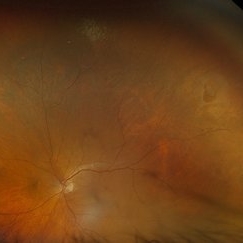
Autofluorescence image of the right eye of a 53-year-old male with history of rheumatic fever and low vision in OU with diagnosis of Extensive Macular Atrophy with Pseudodrusen (EMAP). The image shows a large area of macular atrophy associated with pseudodrusen. Genetic testing was negative for hereditary retinal diseases.
Photographer: Gabriel Andrade
Condition/keywords: Autoflourescence, macula, Retina
-
 Dystrophy of the Retinal Pigment Epithelium
Aug 21 2025 by Aditya S Kelkar, MS, FRCS, FASRS,FRCOphth
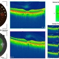
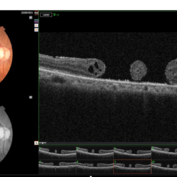
Right eye OCT of a 56 year old female with complaints of gradual painless blurring of vision, aggravating on near work.
Photographer: Dr. Muskan Mangal
Condition/keywords: Dystrophy of the Retinal Pigment Epithelium
-
 Myelinated Nerve Fiber Layer
Aug 13 2025 by Kimberly Wakester
Optomap RGB of a 3-year-old girl that presents with extensive myelinated never fiber in the right eye sparing the fovea. Patient is to return in 6 months for follow up visit with repeat Optos imaging.
Photographer: Kimberly Wakester, COA, OCT-C, Retina Consultants of Carolina
Imaging device: Optos California
Condition/keywords: myelinated nerve fiber layer
-
 Commotio Retinae
Aug 7 2025 by Gabriel Costa Andrade, PhD
Color fundus photograph of a 13-year-old girl who was hit by accidental discharge of gel bullet in her right eye. She presented with retinal whitening with intraretinal hemorrhages in temporal inferior area of the peripheral retina.
Photographer: Gabriel Andrade
Condition/keywords: macula, Retina, Trauma
-
 Macular Star
Aug 6 2025 by Tadeo Blanco
Fundus photograph of a 44 year-old woman with a macular star. She has type 2 diabetes, close living with 8 cats. Presents with decreased visual acuity in the right eye with 1 month of evolution. Refers a febrile episode one month prior. Visual acuity: right eye 20/200, left eye 20/25. Ishihara test: right eye 4/10, left eye 10/10. Bartonella henselae IgG: positive.
Photographer: R. Tadeo Blanco-Nunez
Condition/keywords: macular star, ocular bartonellosis
-
 Horseshoe Retinal Tear
Aug 6 2025 by Korey Starkey
80 year-old patient presented with HSRT without detachment in the left eye and macula-off detachment in the right eye. Scheduled patient for prompt surgical repair OD and same day laser retinopexy OS to reduce risk of retinal detachment.
Photographer: Korey Starkey
Imaging device: Optos
Condition/keywords: color fundus photograph, fundus photography, horseshoe tear, Optos
-
Vortex Vein Varix
Jul 29 2025 by Gordon Crabtree, MD
Right eye B-scan video of 41-year-old man with bilateral vortex vein vortices demonstrating reduction in elevation of varix elevation from 0.92mm to flat with prolonged upgaze.
Condition/keywords: vortex vein varix
-
Subhyaloid Hemorrhage
Jul 14 2025 by SHRADDHA ASHOK CHANDORKAR, DNB DO FVRS
19 year old female presented with sudden blurring of vision in her right eye since few hours after she attended a DJ party the previous night. On examination Vision was counting fingers close to face and Retina showed Subhyaloid hemorrhage with some RPE damage. YAG hyaloidotomy was performed and the subhyaloid hemorrhage was drained. Need for injections if RPE damage and development of CNV in future was explained. Patient was apprehensive as the vision was not restored immediately after the blood was drained. On subsequent follow ups slowly patient’s vision was restored to 6/6N6 after about a month.
Condition/keywords: subhyaloid hemorrhage
-
 Subhyaloid Hemorrhage
Jul 12 2025 by SHRADDHA ASHOK CHANDORKAR, DNB DO FVRS
19 year old female presented with sudden blurring of vision in her right eye since few hours after she attended a DJ party the previous night. On examination Vision was counting fingers close to face and Retina showed Subhyaloid hemorrhage with some RPE damage. YAG hyaloidotomy was performed and the subhyaloid hemorrhage was drained. Need for injections if RPE damage and development of CNV in future was explained. Patient was apprehensive as the vision was not restored immediately after the blood was drained. On subsequent follow ups slowly patient’s vision was restored to 6/6N6 after about a month.
Photographer: Dr.Shraddha Chandorkar
Imaging device: Zeiss
Condition/keywords: subhyaloid hemorrhage
-
 Prepapillary Vascular Loop
Jul 4 2025 by KANWALJEET HARJOT MADAN, M.S. (Ophthalmology); FAICO (Vitreous - Retina)
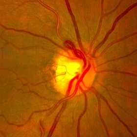
This is the fundus picture of right eye of a young 32 years female depicting pre papillary vascular loop. A prepapillary vascular loop is a congenital anomaly of the optic disc that presents as an elevated and twisted bundle of vessels projecting into the vitreous cavity. It is a benign condition, usually unilateral but can be bilateral. It is asymptomatic and discovered during routine eye examination. This anomaly can sometimes cause complications like branch retinal artery occlusion, vitreous hemorrhage, or sub retinal hemorrhage.
Photographer: Dr. Kanwaljeet Harjot Madan, Thind Eye Hospital, Jalandhar City (Punjab) INDIA.
Imaging device: Zeiss Fundus Camera
Condition/keywords: branch retinal artery occlusion (BRAO), optic disc, Prepapillary Vascular Loop, SUB RETINAL HEMORRHAGE, Vitreous hemorrhage
-
 Fluorescein Angiography (FA) of a Primary Retinal Vasoproliferative Tumor
Jun 29 2025 by Marcelo Zas, MD PhD
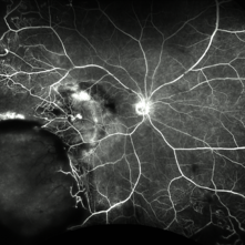
We present a case of a 33-year-old male patient, who presented with decreased visual acuity in his right eye with 20/80, presenting a primary retinal vasoproliferative tumor in the lower temporal quadrant. The fluorescein angiography findings are: 1. Early hyperfluorescence due to its rich intrinsic vascularity and often has dilated feeding arterioles and draining venules. 2. Marked progressive leakage from the tumor vessels. 3. The late leakage often obscures fine vascular details in the late phase and corresponds to exudation and macular edema seen clinically. 4. Staining of surrounding exudates, RPE disturbances and gliosis. 5. In our case also a marked peripheral capillary closure in the areas adjacent to the tumor and in other quadrants as well.
Photographer: Marcelo Zas MD PhD
Condition/keywords: RETINAL VASOPROLIFERATIVE TUMOR
-
 Fluorescein Angiography in Choroidal Rupture
Jun 26 2025 by Hector Gabriel Moreno Solano, MD, MHA
Fluorescein angiography of the right eye reveals three linear hypofluorescent lesions with progressive staining at the edges, consistent with choroidal ruptures. These lesions are temporally located in the posterior pole, with one of them situated near the fovea but without direct foveal involvement. The pattern is suggestive of previous blunt ocular trauma.
Photographer: Héctor Gabriel Moreno Solano, Instituto Mexicano de Oftalmología “IMO I.A.P”
Imaging device: CLARUS
Condition/keywords: Choroidal Rupture, fluorescein angiogram (FA)
-
 OCT Choroidal Rupture
Jun 26 2025 by Hector Gabriel Moreno Solano, MD, MHA
High-resolution OCT of the right eye shows a localized disruption of the retinal pigment epithelium (RPE)–Bruch’s membrane complex, consistent with a choroidal rupture. There is loss of the normal outer retinal architecture over the lesion, with focal elevation and irregularity of the underlying RPE. Hyperreflective material is noted at the level of the break, without associated subretinal fluid or signs of active choroidal neovascularization.
Photographer: Hector Gabriel Moreno Solano, Instituto Mexicano de Oftalmología “IMO I.A.P”
Imaging device: REVO
Condition/keywords: Choroidal Rupture, OCT
-
 Autofluorescence in Multiple Choroidal Ruptures
Jun 26 2025 by Hector Gabriel Moreno Solano, MD, MHA
Fundus autofluorescence imaging of the right eye shows three hypoautofluorescent linear lesions located temporally to the fovea, consistent with choroidal ruptures. The lesions demonstrate sharply demarcated borders with variable surrounding hyperautofluorescence, suggestive of retinal pigment epithelium (RPE) disruption and potential remodeling. One rupture is located near the foveal region, though the foveal center remains spared.
Photographer: Hector Gabriel Moreno Solano, Instituto Mexicano de Oftalmología “IMO I.A.P”
Imaging device: CLARUS
Condition/keywords: autofluorescence imaging, Choroidal Rupture
-
 Multiple Chorodial Ruptures
Jun 26 2025 by Hector Gabriel Moreno Solano, MD, MHA
Color fundus photograph of the right eye reveals three well-defined, curvilinear choroidal ruptures located temporal to the fovea running parallel. The lesions appear as pale, crescent-shaped bands, with underlying retinal pigment epithelium disruption. One of the ruptures is situated near the foveal center, though without direct involvement.
Photographer: Hector Gabriel Moreno Solano, Instituto Mexicano de Oftalmología “IMO I.A.P”
Imaging device: CLARUS
Condition/keywords: Choroidal Rupture, color fundus photograph, color wide field
-
 Double Macular Holes
Jun 26 2025 by Moazzam Parvez
OCT image of a 62 year old man after a blunt trauma by a tennis ball with a vision of CF 3 mt in the right eye.
Photographer: Moazzam Parvez , Netralayam , Kolkata
Imaging device: Topcon Maestro 2
Condition/keywords: double, traumatic macular hole
-
 Two Suns in the Macular Sky
Jun 26 2025 by Moazzam Parvez
Fundus photograph of a 62 year old gentleman presenting with double adjacent full thickness macular holes in the right eye maintaining a vision of CF 3 mts.
Photographer: Moazzam Parvez ,Netralayam , Kolkata
Imaging device: Topcon Maestro 2
Condition/keywords: double, Macular hole, traumatic macular hole
-
 Epiretinal Membrane
Jun 25 2025 by Kimberly Wakester
Fundus photograph of a 32-year-old woman with a stable epiretinal membrane in the right eye. Patients vision remains stable. No intervention is required at this time.
Photographer: Kimberly Wakester, COA, OCT-C
Imaging device: Topcon TRC 50DX
Condition/keywords: ERM
-
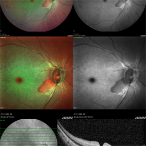 CRAO With Cilio-retinal Sparing-MMI
Jun 25 2025 by Shivankar Sen, MS, FVRS
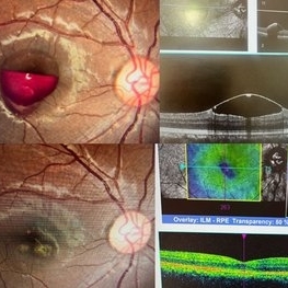
A 41 year old male came with complaints of Right eye blurring of vision since a day associated with watering and redness. He had no systemic illness, though gave a history of fall from bike 1 month back at the time of which he had blunt force trauma to the right side of the face. BCVA was 3/60, less than N36 in the right eye and 6/6, N6 in the left eye. Right eye had Marcus Gunn Pupil with clear lens, Left eye was within normal limits. IOP was normal; 16 in OD and 18 in OS. Retina evaluation revealed CRAO in the right eye with cilio-retinal artery sparing. Left eye was unremarkable Image Details Left to Right (Top 2 rows) Multicolor Reflectance Image (Blue-green enhanced 55 degree) revealing cilioretinal spared retinal stroma and a characteristic Cherry Red Spot; Green Reflectance showing corresopnding dark gray area with spared perfusion and black spot consistent with Cherry Red Spot on multicolor 2nd Row - 35 degree image (Multicolor Standard Reflectance and Green Reflectance) 3rd Row - SD-OCT revealing acute moderate CRAO findings with Middle retinal layer opacification and prominent middle limiting membrane (p-MLM) sign; Inner retinal layer opacification and prominent retinal pigment epithelium at the fovea with Diminished inner retinal layer stratification
Photographer: Gayathri M S
Imaging device: Heidelberg Spectralis HRA+OCT
Condition/keywords: CRAO with cilioretinal sparing, multicolor, multimodal imaging, OCT biomarkers, reflectance
-
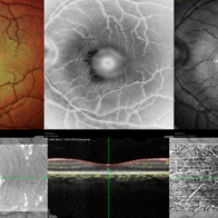 Berlins Edema - Multimodal Imaging
Jun 25 2025 by Shivankar Sen, MS, FVRS
A 22 year old female came with history of injury to her left eye with a badminton racquet butt cap an hour before presentation On examination, she was found to have right eye 6/6;N6 vision and within normal limits, left eye 6/9;N6 vision, cells1+ in the anterior chamber, brisk pupillary response, no vitreous reaction and sub-clinical berlin's edema at the posterior pole. Multimodal imaging revealed frank boundaries of Berlin's edema more pronounced in the nasal parafoveal region. Figure details Top (Left to Right) Multicolor Reflectance showing bright yellow ring surrounding the perifovea; Blue Reflectance (Black on white contrast) showing corresponding black ring; Green Reflectance showing a characteristic white ring (all pronounced nasally); Bottom (Left-Right) Transverse structural OCT enface image showing white ring consistent with edema OCTA inner layer segmentation from ILM to GCL Transverse corresponding OCTA revealing faint hypo ring within perifoveal capillary bed
Photographer: Gayathri M S
Imaging device: Heidelberg Spectralis HRA+OCT
Condition/keywords: blue reflectance, En Face OCTA, enface imaging, multicolor, oct, reflectance
-
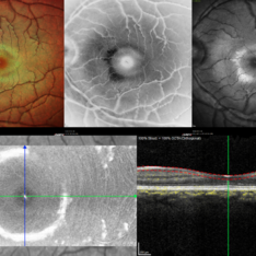 Berlins
Jun 25 2025 by Shivankar Sen, MS, FVRS
A 22 year old female came with history of injury to her left eye with a badminton racquet butt cap an hour before presentation On examination, she was found to have right eye 6/6;N6 vision and within normal limits, left eye 6/9;N6 vision, cells1+ in the anterior chamber, brisk pupillary response, no vitreous reaction and sub-clinical berlin's edema at the posterior pole. Multimodal imaging revealed frank boundaries of Berlin's edema more pronounced in the nasal parafoveal region. Figure details Top (Left to Right) Multicolor Reflectance showing bright yellow ring surrounding the perifovea; Blue Reflectance (Black on white contrast) showing corresponding black ring; Green Reflectance showing a characteristic white ring (all pronounced nasally); Bottom (Left-Right) Transverse structural OCT enface image showing white ring consistent with edema OCTA inner layer segmentation from ILM to GCL
Photographer: Gayathri M S
Imaging device: Heidelberg Spectralis HRA+OCT
Condition/keywords: blue reflectance, En Face OCTA, multicolor
-
 Retinal Vasoproliferative Tumor
Jun 24 2025 by Marcelo Zas, MD PhD
We present a case of a 33-year-old male patient, who presented with decreased visual acuity in his right eye with 20/80, presenting a primary retinal vasoproliferative tumor in the lower temporal quadrant. The tumor is associated with serous retinal detachment, hard exudation, neovascularization and telangiectasias. Lipid exudates extend toward the macula, indicating macular involvement, which may contribute to decreased visual acuity. Oi was normal with 20/20 of BCVA. The patient was treated initially with IV anti-VEGF therapy and cryotherapy.
Photographer: Marcelo Zas MD PhD
Condition/keywords: RETINAL VASOPROLIFERATIVE TUMOR
-
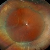 Repaired Retinal Detachment
Jun 24 2025 by Kimberly Wakester
Optomap RGB of an 45-year-old woman with a repaired retinal detachment in the right eye. The operative eye is doing well three-month s/p surgery. Retina is attached 360 on SB. There is resolving residual SRF at 6:00. Discussed the possible need for added laser. Will continue to observe and will return in 3 months for follow up exam.
Photographer: Kimberly Wakester, COA, OCT-C
Imaging device: Optos California
Condition/keywords: repaired RD, scleral buckle
 Loading…