 Initializing download.
-
 By Pablo Angel Garcia Uribe
Clínica Oftalmológica Salauno
Co-author(s): Optom. Marilyn Alvarez Monroy, Clínica Oftalmológica Salauno - Uploaded on Sep 3, 2025.
- Last modified by Joshua Friedman on Sep 4, 2025.
- Rating
- Appears in
- Miscellaneous
- Condition/keywords
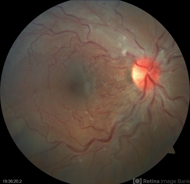
- papillophlebitis
- Photographer
- Clínica Oftalmológica Salauno
- Imaging device
-
Fundus camera
Visucam 524, Carl Zeiss Meditec AG, Jena, Germany - Description
- Fundus photograph of a 24-year-old woman, previously healthy, with a history of recreational inhaled cannabis use, presented with a 24-hour history of photopsias and mild decrease in visual acuity, associated with a subtle relative central scotoma in the right eye. On ophthalmic examination, the anterior segment of both eyes was unremarkable. Best-corrected visual acuity was slightly reduced in the right eye and normal in the left. Fundus biomicroscopy of the right eye revealed moderate disc edema with hyperemia and well-defined margins, accompanied by venous engorgement and tortuosity, predominantly affecting the venules. No retinal hemorrhages were observed. Additionally, retinal thickening was noted along the temporal arcades, with apparent foveal sparing. The left eye showed no pathological findings. Based on the patient’s age, the acute onset of symptoms, the fundoscopic features, and the absence of systemic risk factors, the clinical presentation was consistent with papillophlebitis.


