 Initializing download.
-
 By Marcelo Zas, MD PhD
Hospital de Clinicas-University of Buenos Aires
Co-author(s): Guido Bregliano MD, Mariano Cotic MD, Julieta Fourcade MD, Marcos Mendaro MD, Maria Carolina Pozzoni MD, Adriana Nieva MD, Luciano Scorsetti MD, Daniela Contartese MD, Sofia Ghigliotti MD, Pablo Chiaradia MD PhD - Uploaded on Jun 29, 2025.
- Last modified by Joshua Friedman on Jun 30, 2025.
- Rating
- Appears in
- Miscellaneous
- Condition/keywords
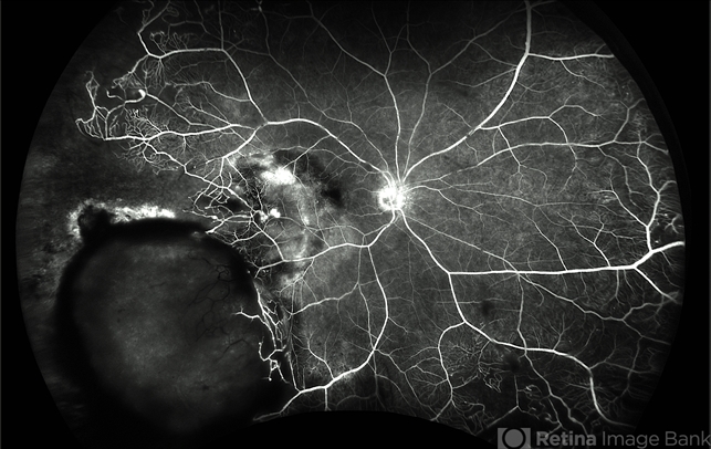
- RETINAL VASOPROLIFERATIVE TUMOR
- Photographer
- Marcelo Zas MD PhD
- Description
- We present a case of a 33-year-old male patient, who presented with decreased visual acuity in his right eye with 20/80, presenting a primary retinal vasoproliferative tumor in the lower temporal quadrant. The fluorescein angiography findings are: 1. Early hyperfluorescence due to its rich intrinsic vascularity and often has dilated feeding arterioles and draining venules. 2. Marked progressive leakage from the tumor vessels. 3. The late leakage often obscures fine vascular details in the late phase and corresponds to exudation and macular edema seen clinically. 4. Staining of surrounding exudates, RPE disturbances and gliosis. 5. In our case also a marked peripheral capillary closure in the areas adjacent to the tumor and in other quadrants as well.

---thumb.jpg/image-square;max$79,0.ImageHandler "Myopic Giant Tear")

---thumb.jpg/image-square;max$79,0.ImageHandler "Retinectomy With Diathermy in a Giant Tear")


