Search results (27 results)
-
 Look With Your Heart
Sep 20 2024 by Virginia Gebhart
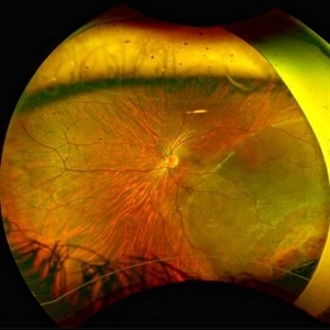
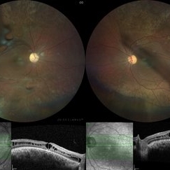
FA of 65 year old male with exudative AMD superior to a chorioretinal defect in the nasal macula. FA shows classic CNV with late leakage. Treated with IVA, will consider PDT if no improvement.
Photographer: Virginia Gebhart, Retina Consultants of Carolina
Imaging device: Optos California
Condition/keywords: choroidal neovascularization (CNV), exudative age-related macular degeneration, FA early phase
-
 Rhegmatogenous retinal detachment in a Young Patient
May 18 2023 by Jesus Lozano, MD
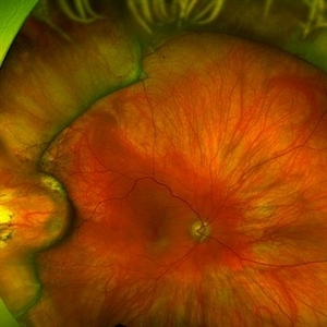
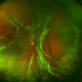
25 year old male patient. High Myope. Arrived to my clinic with a VA Fc 2mts. - Optos Image : Rhegmatogenous Retinal Detachment Mácula Off with ínfero temporal retinal holes in an area of lattice degeneration. Visual Acuity FC 2mts.
Imaging device: Optos
Condition/keywords: retinal detachment with retinal defect
-
 Giant Retinal Tear
Mar 15 2022 by Jesus Lozano, MD
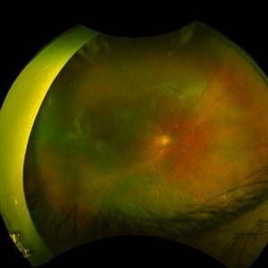
42 year old woman with a supero temporal giant retinal tear.
Photographer: Dr. Mohamad Midlij
Imaging device: Optos Silverstone
Condition/keywords: retinal detachment with retinal defect, retinal tear
-
 Scleral Buckle Surgery
Mar 14 2022 by Jesus Lozano, MD
27 year old man after 1 Week Scleral Buckle Surgery. 4.0mm Silicon Strip. Retina attached.
Photographer: Dr. Avi Schwalb, Hillel Yaffe Medical Center, Israel.
Imaging device: Optos Silverstone
Condition/keywords: retina surgery, retinal detachment with retinal defect, scleral band, scleral buckle
-
 Rhegmatogenous retinal detachment in a 27 year old man.
Mar 14 2022 by Jesus Lozano, MD
27 year old man with a rhegmatogenous retinal detachment macula off.
Photographer: Dr. Avi Schwalb, Hillel Yaffe Medical Center, Israel.
Imaging device: Optos Silverstone
Condition/keywords: retina surgery, retinal detachment with retinal defect, scleral band, scleral buckle
-
 Erosion of Segmental Buckle
Feb 25 2022 by Roger A. Goldberg, MD, MBA
Erosion of sharp edge of segmental scleral buckle seen 15 years after being placed for repair of a retinal detachment
Photographer: Melissa Bartlett, Bay Area Retina Associates
Imaging device: Optos
Condition/keywords: retinal defect, scleral buckle
-
 Retinal Detachment with Proliferative Vitreoretinopathy
Jan 31 2022 by Ahmad B. Tarabishy, MD
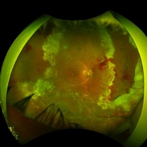
Ultra wide-field fundus photograph of a 55-year-old gentleman who had previously underwent laser retinopexy for multiple inferior retinal breaks. He presented with a macula-off retinal detachment from a new temporal break with proliferative vitreoretinopathy with fixed folds noted temporally and superonasally.
Photographer: Megan McLandsborough, Lakeland Eye Clinic
Imaging device: Optos California UWF Camera
Condition/keywords: laser retinopexy, macula off Retinal Detachment, proliferative retinopathy, proliferative vitreoretinopathy (PVR), Retinal Detachment, retinal detachment with retinal defect
-
 Retinal Detachment Following Scleral Buckling, Retinectomy, Laser, and Oil
Jan 31 2022 by Ahmad B. Tarabishy, MD
Ultra wide-field fundus photograph of a 55-year-old gentleman who is 4 days after surgery with scleral buckling, pars plana vitrectomy, perfluoron tamponade, membrane peeling, direct fluid-PFO-oil exchange, nasal and temporal retinectomies, and endolaser photocoagulation. Visual acuity was 20/150 under oil.
Photographer: Megan McLandsborough, Lakeland Eye Clinic
Imaging device: Optos California UWF Camera
Condition/keywords: endolaser, Membrane Peel, PPV, proliferative retinopathy, proliferative vitreoretinopathy (PVR), Retinal Detachment, retinal detachment with retinal defect, scleral buckle, submacular perfluorocarbon liquid (PFO)
-
 Retinal Detachment with Tear
Jan 11 2022 by Manish Nagpal, MD, FRCS (UK), FASRS
Intraoperative photo of a retinal detachment with a posterior tear with everted edges.
Photographer: Manish Nagpal, Retinal Foundation, Ahmedabad, India
Imaging device: Sony PMW -10 MD surgical camera
Condition/keywords: retinal detachment of the macula, retinal detachment with retinal defect, retinal detachment with tear
-
 Rhegmatogenous Retinal Detachment in Myopic Patient
Sep 19 2021 by Jesus Lozano, MD
Rhegmatogenous Retinal Detachment in a 50 year-old man with high myopia.
Photographer: Yair Bet Yosef, Hadassah Medical Center. Israel
Imaging device: Optos Silverstone
Condition/keywords: high myopia, retinal detachment with retinal defect
-
 Macular Hole Retinal Detachment
Sep 14 2021 by Ogugua Ndubuisi Okonkwo, MD, FRCS (Edin), FASRS
Preoperative optical coherence tomogram (OCT) of the right eye in a 65-year-old male. This shows a detached retina with significant subretinal fluid in the macular area. There is a full-thickness defect and discontinuity of the foveomacula, which represents a detached macular hole.
Photographer: Oreoluwa Olabode , Eye Foundation Hospital, Lagos.
Imaging device: Optovue Avanti RTVue.
Condition/keywords: macular hole retinal detachment, retinal detachment with retinal defect
-
 Retinal Detachment with Giant Tear
Jul 6 2021 by Lucas Zago Ribeiro, MD
Fundus composite of an 50-year-old man with macula-off retinal detachment due to temporal giant retinal tear.
Photographer: Lucas Zago Ribeiro, Federal University of São Paulo (UNIFESP)
Imaging device: Zeiss Visucam 524
Condition/keywords: giant retinal tear, retinal detachment with retinal defect
-
 Total Rhegmatogenous Retinal Detachment With Horseshoe Tear and PVR
Jun 19 2021 by Kushal S Delhiwala, MBBS, MS, FMRF,FICO, FAICO
Ultra-widefield fundus photograph of an 60-year-old pseudophakic female showing total rhegmatogenous retinal detachment and horse shoe retinal tear temporally along with circumferential PVR changes and star folds.
Photographer: Kushal Delhiwala, Netralaya superspeciality eye hospital, Ahmedabad, Gujarat,India
Imaging device: Optos Daytona
Condition/keywords: proliferative vitreoretinopathy (PVR), retinal detachment with retinal defect, star folds, ultra-wide field imaging
-
 Juvenile X-Linked Retinoschisis
Mar 4 2021 by SHISHIR VERGHESE, MS, FVRS, FAICO (Retina)
Fundus Photograph of a 25-year-old male with juvenile x-linked retinoschisis with inner retinal defects seen above superior and inferior arcades in both eyes. Corresponding OCT showing retinal schisis involving fovea.
Photographer: Shishir Verghese, Aravind Eye Hospital and Postgraduate institute of Ophthalmology, Coimbatore
Imaging device: Zeiss Clarus
Condition/keywords: juvenile retinoschisis, retinoschisis
-
 Macula ON Retinal Detachment
Feb 18 2021 by Omar Lezrek
Fundus mosaic of an 26-year-old woman with macula ON retinal detachment with 9 o'clock retina tear.
Photographer: Omar Lezrek MD
Imaging device: Eidon AF
Condition/keywords: retinal detachment with retinal defect
-
 Retinal Detachment
Aug 23 2020 by Renata Bertazzi
Fundus photograph of an 66-year-old man with retinal detachment with superior temporal horseshoe rupture
Photographer: Renata Bertazzi, Instituto Paulista de ensino e Pesquisa em Oftalmologia, São Paulo, SP
Imaging device: DayTona - Optos
Condition/keywords: retinal detachment with retinal defect
-
 Surfboard to OS - Choroidal Rupture, Retinal and Choroidal Avulsion, Retinal Detachment
May 20 2020 by Theodore Leng, MD, MS, FASRS
33-year-old male who sustained a surfboard to the left orbit with orbital fractures, choroidal rupture, nasally and retinal and choroidal avulsion nasally. He also had a horseshoe retinal tear superotemorally and a resultant rhegmatogenous retinal detachment.
Condition/keywords: choroidal avulsion, choroidal rupture, retinal avulsion, retinal detachment with retinal defect
-
 Coats' Related Total Serous Retinal Detachment
Jan 19 2020 by Anfisa Ayalon, MD
Slit-lamp photograph of a 3 -year-old male with total serous retinal detachment due to Coats' disease in the right eye. S/p laser photocoagulation, cryotherapy, retinal detachment repair with scleral buckle implantation 2 years ago. Currently, the right eye has no light perception.
Photographer: Anfisa Ayalon, MD., Meir Medical Center, Kfar Saba, Israel.
Condition/keywords: blind eye, Coats' disease, retinal detachment without retinal defect, serous retinal detachment
-
 Retinal Detachment and Inferonasal Dialysis
Nov 27 2018 by Maria H. Berrocal, MD
66-year-old who suffered blunt trauma to the eye and presented with inferonasal dialysis and retinal detachment.
Photographer: Thaylan Calderon, Berrocal & Associates, San Juan, PR
Imaging device: Optos
Condition/keywords: retinal detachment with retinal defect
-
 Asymptomatic Superior Retinal Detachment
May 5 2016 by Steven J Ryder, MD
38-year-old African American female with moderate myopia (-4.50 Sph OU) and asymptomatic superior retinal detachment in the right eye. Zeiss OCT capturing vertical raster scans through border of retinal detachment.
Photographer: Luis Bernhard, Miami VA Healthcare System
Imaging device: Cirrus
Condition/keywords: asymptomatic, full thickness retinal hole, retinal detachment with retinal defect
-
 Asymptomatic Superior Retinal Detachment
May 5 2016 by Steven J Ryder, MD
38-year-old African American female with moderate myopia (-4.50 Sph OU) and asymptomatic superior retinal detachment in the right eye. Zeiss Cirrus OCT capturing full-thickness retinal break at 12:00 and temporal vitreoretinal traction.
Photographer: Luis Bernhard, Miami VA Healthcare System
Imaging device: Zeiss Cirrus
Condition/keywords: asymptomatic, full thickness retinal hole, retinal break, retinal detachment with retinal defect
-
 Asymptomatic Superior Retinal Detachment
May 5 2016 by Steven J Ryder, MD
38-year-old African American female with moderate myopia (-4.50 Sph OU) and asymptomatic superior retinal detachment in the right eye. Montage fundus photography showing localized retinal detachment superiorly with single full-thickness retinal break at 12:00.
Photographer: Luis Bernhard, Miami VA Healthcare System
Imaging device: Topcon
Condition/keywords: asymptomatic, full thickness retinal hole, myopia, retinal break, retinal detachment with retinal defect
-
 Funnel Retinal Detachment
Feb 2 2015 by Matt Poe, COA
The patient presented with total vision loss for >2months. Patient had history of exudative ARMD with intravitreal injections. No surgical intervention was done due to the long standing detachment and patient health.
Photographer: Matt Poe, COA. Northwest Arkansas Retina Associates, Springdale, AR.
Condition/keywords: retinal defect
-
 Retinal Detachment with Proliferative Vitreoretinopathy
Mar 20 2014 by Min Kim, MD, PhD, MBA, FASRS
Wide field fundus photograph of a 59-year-old male with chronic total RD and PVR, with multiple retinal breaks that developed a few months after LASIK surgery.
Photographer: Young Duk Bae, Yonsei University, Gangnam Severance Hospital
Imaging device: Wide field fundus photography, Optomap
Condition/keywords: proliferative vitreoretinopathy (PVR), retinal detachment with retinal defect
-
---thumb.JPG/image-square;max$300,300.ImageHandler) Retinal Detachment
Retinal Detachment
Jul 13 2013 by Jason S. Calhoun
Fundus photo shows retinal detachment.
Photographer: Jason S. Calhoun, Department of Ophthalmology, Mayo Clinic Jacksonville, Florida
Condition/keywords: retinal detachment with retinal defect
 Loading…