Search results (49 results)
-
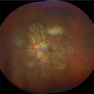
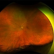
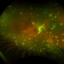
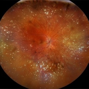 VKH Syndrome
Jun 12 2025 by Virginia Gebhart
22 year old male with VKH Syndrome. Pt has been experiencing severe headaches, distorted vision, hearing loss, weakness, and a large white patch of hair. Significant cell in AC and vitreous, multiple punched-out CR scars in periphery. Referred to rheumatology for possible immunomodulatory treatment
Photographer: Virginia Gebhart, Retina Consultants of Carolina
Imaging device: Optos California
Condition/keywords: montage, multifocal choroiditis, panuveitis, Vogt-Koyanagi-Harada
-
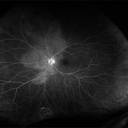
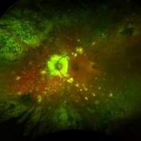
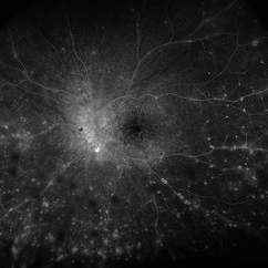 VKH Syndrome
Jun 12 2025 by Virginia Gebhart
Fluorescein angiogram of 22 year old male with VKH syndrome. Significant cell in AC and vitreous, multiple punched-out CR scars in periphery, mild vascular leakage. Pt referred to rheumatology for immunomodulatory treatment.
Photographer: Virginia Gebhart, Retina Consultants of Carolina
Imaging device: Optos California
Condition/keywords: FA, fluorescein angiogram (FA), multifocal choroiditis, panuveitis, VKH, Vogt-Koyanagi-Harada
-
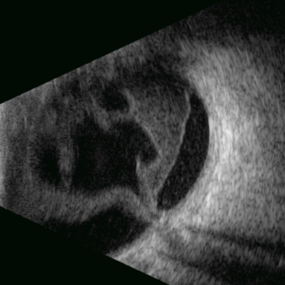 Stag Horn
Apr 8 2025 by Gustavo Uriel Fonseca Aguirre
B-mode ultrasound of a young male patient with bilateral panuveitis (currently under investigation) reveals intense vitritis with islands of preserved vitreous and partial posterior hyaloid detachment, creating a characteristic "stag horn" appearance.
Photographer: Gustavo U. Fonseca Aguirre, Hospital Conde de Valenciana, Ciudad de México
Condition/keywords: Panuveitis
-
 Panuveitis
Jul 12 2024 by Korey Starkey
Ultra widefield Optos FA of 59 year old female presents with panuveitis in both eyes. Patients vision was VA OS: Dcc20/60-2 at time of visit.
Photographer: Korey Starkey
Imaging device: Optos
Condition/keywords: FLUORESCEIN ANGIOGRAPHY, hyperfluorescence, Optos, Panuveitis, ultra-wide field imaging, Uveitis
-
 Atypical Tubercular Peripheral Occlusive Retinal Vasculitis
Jun 21 2024 by Tejaswita Verma
Fundus montage of the right eye of a 27 year old male with macula threatening occlusive vasculitis showing hemorrhages in inferior, temporal quadrant with vascular sheathing. The patient was Mantoux positive (20 mm induration) and IGRA (TB-GOLD)positive and started on oral steroids. The case was atypical due to no vitritis at presentation which is unusual of tuberculosis. Behcet's disease was ruled out as there was no panuveitis like picture.
Photographer: DR. TEJASWITA VERMA
Imaging device: MIRANTE
Condition/keywords: occlusive vasculitis, ocular tuberculosis
-
 Panuveitis
Apr 2 2024 by Zach Seim
Optos Ultra-widefield photo OS of a 59 year old female with Panuveitis OU.
Photographer: Zach Seim
Imaging device: Optos California
Condition/keywords: Optos, OPTOS CALIFORNIA, panuveitis, ULTRA WIDE FIELD, ultra-wide field imaging
-
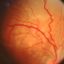
 VKH - Uveitic stage
Jun 23 2023 by Sergio Emilio Sifuentes Renteria, MD
Fundus photograph of a young female with VKH in uveitic stage
Photographer: Sergio Emilio Sifuentes Rentería - Foundation Hospital Nuestra Señora de La Luz
Condition/keywords: choroiditis, panuveitis, serous retinal detachment, VKH, Vogt-Koyanagi-Harada
-
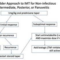 Posterior Manifestations of Sarcoidosis – Management of noninfectious intermediate, posterior, or panuveitis
Mar 29 2023 by Joshua Friedman
Management of noninfectious intermediate, posterior, or panuveitis.
Condition/keywords: panuveitis, sarcoidosis
-
 Vogt-Koyanagi-Harada Disease
Apr 24 2022 by Aniruddha K Agarwal, MD
A 38-year-old woman of Asian descent with no ophthalmological or systemic history presented to the emergency eye clinic with a 1-week complaint of headache and bilateral vision loss. Funduscopy revealed bilateral serous neurosensory detachments. The presence of lymphocytosis in cerebrospinal fluid and mild acute sensorineural hearing loss confirmed the diagnosis of uveomeningoencephalitic syndrome (Vogt-Koyanagi-Harada disease).
Photographer: Mercedes SERRADOR, MD, PhD and Beatriz VENTAS, MD
Imaging device: Zeiss Clarus fundus camera
Condition/keywords: IUSG, panuveitis, Vogt-Koyanagi-Harada
-
 Multifocal Choroiditis and Panuveitis- Schlaegel lines
Nov 16 2021 by Manuel Ángel Alcántara Delgado, MD
Optomap ultra-widefield retinal imaging of an 52-year-old woman showed multiple punched-out chorioretinal lesions and 2 rows of peripheral curvilinear pigmented chorioretinal streaks (Schlaegel lines).
Photographer: Manuel Ángel Alcántara Delgado. Conde de Valenciana.
Condition/keywords: multifocal choroiditis, myopia, retina, uveitis
-
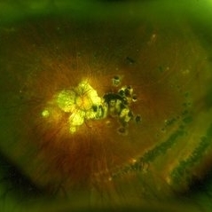
 Pigmented Vitreous Cyst
May 25 2021 by Anmol Naik
A 56-year-old Indian male, known case of panuveitis, presented with complaint of floater in his left eye. On examination, a pigmented floating vitreous cyst was seen in the anterior vitreous. Vitreous cyst, an extremely rare entity, can be congenital or acquired. Detailed examination is necessary to rule out malignancy and parasitic infections.
Photographer: Dr. Anmol Naik, MS, Nakshatra Superspeciality Eye Hospital, Pune, India
Imaging device: Appasamy Associates Slit Lamp AIA-11 3S L model, Chennai, India
Condition/keywords: vitreous cyst
-
 Serous Retinal Detachment in Vogt Koyanagi Harada Patient
Apr 26 2021 by Pablo Baquero Ospina, MD
24-year-old woman with bilateral panuveitis and serous retinal detachment, headache and tinnitus.
Photographer: Pablo Baquero-Ospina, Asociación Para Evitar la Ceguera en México
Imaging device: Heidelberg Spectralis
Condition/keywords: serous retinal detachment, Vogt-Koyanagi-Harada
-
 Acute Syphilitic Posterior Placoid Chorioretinitis
Nov 22 2020 by Shawn Sell
58-year-old homeless male presenting with 2 weeks of bilateral eye redness and photosensitivity found to have panuveitis with a positive VDRL CSF and RPR titer of 1:512 with acute syphilitic posterior placoid chorioretinitis.
Photographer: Eastern Virginia Medical School
Imaging device: Optos
Condition/keywords: acute syphilitic posterior placoid chorioretinitis, neurosyphilis
-
 Acute Syphilitic Posterior Placoid Chorioretinitis
Nov 22 2020 by Shawn Sell
58-year-old homeless male presenting with 2 weeks of bilateral eye redness and photosensitivity found to have panuveitis with a positive VDRL CSF and RPR titer of 1:512 with acute syphilitic posterior placoid chorioretinitis.
Photographer: Eastern Virginia Medical School
Imaging device: Optos
Condition/keywords: acute syphilitic posterior placoid chorioretinitis
-
 Multifocal Choroiditis and Panuveitis Scars
Sep 4 2020 by Harvey S Uy, MD
56-year-old female with 10 year history of recurrent mild uveitis and increasing choroidal scars.
Condition/keywords: multifocal chorioretinitis (MCP)
-
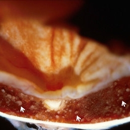 Vogt-Koyanagu-Harada (VKH) Disease
May 18 2020 by McGill University Health Centre
VKH disease is an autoimmune condition that causes bilateral chronic granulomatous panuveitis, and extraocular manifestations in the central nervous system, auditory system, and integument. VKH disease is twice as prevalent in women than men, and is believed to be associated with specific human leukocyte antigen (HLA) types, suggesting a possible hereditary component. The exact cause of VKH disease, however, remains unclear. Image (B) shows several aggregate RPE cells and histiocytes, called Dalen–Fuchs nodules (arrows)
Condition/keywords: Dalen-Fuchs nodules, enucleation, Vogt-Koyanagi-Harada
-
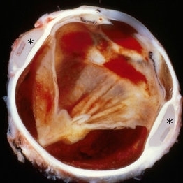 Vogt-Koyanagu-Harada (VKH) Disease
May 18 2020 by McGill University Health Centre
VKH disease is an autoimmune condition that causes bilateral chronic granulomatous panuveitis, and extraocular manifestations in the central nervous system, auditory system, and integument. VKH disease is twice as prevalent in women than men, and is believed to be associated with specific human leukocyte antigen (HLA) types, suggesting a possible hereditary component. The exact cause of VKH disease, however, remains unclear. In (A), an enucleation specimen shows a retinal detachment and a scleral buckle (*).
Condition/keywords: scleral buckle, Vogt-Koyanagi-Harada
-
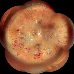
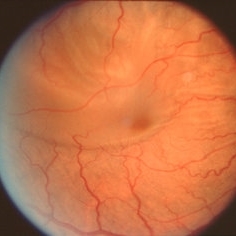
 Resolved Exudative RD in Vogt-Koyanagi-Harada Syndrome
Mar 27 2019 by Gary R. Cook, MD, FACS
Right eye of the same patient following resolution of the exudative retinal detachment OD.
Condition/keywords: exudative detachment, panuveitis, Vogt-Koyanagi-Harada
-
 Vogt-Koyanagi-Harada Syndrome
Mar 27 2019 by Gary R. Cook, MD, FACS
View along superior arcade of exudative retinal detachment secondary to Vogt-Koyanagi-Harada Syndrome OD.
Condition/keywords: exudative detachment, panuveitis, Vogt-Koyanagi-Harada
-
 Vogt-Koyanagi-Harada Syndrome
Mar 27 2019 by Gary R. Cook, MD, FACS
Exudative retinal detachment secondary to Vogt-Koyanagi-Harada syndrome OD
Condition/keywords: exudative detachment, panuveitis, Vogt-Koyanagi-Harada
-
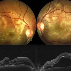 Vogt-Koyanagi-Harada Disease
Feb 13 2019 by Deepak Bhojwani, MS
A 18-year-old girl came with complaints of acute onset of diminution of vision associated with dull boring pain and headache. She gave a history of flu like symptoms with fever headache and malaise few days before onset of ocular symptoms. Fundus photographs shows classic picture of acute stage of VKH with multiple large serous RPE detachments and inflamed choroid. OCT shows hallmark features of VKF viz. multiple serous RPEDs with hyperreflective dots, subretinal septa and thick choroid.
Photographer: DR DEEPAK BHOJWANI
Imaging device: Zeiss Visucam 500
Condition/keywords: bilateral serous detachment, panuveitis, Vogt-Koyanagi-Harada
-
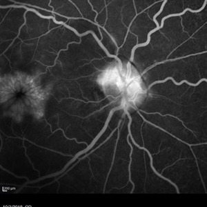 Weiss Ring
Jan 15 2019 by Olivia Rainey
Fluorescein angiogram of a 55-year-old female with a Weiss ring affecting her right eye. Patient was diagnosed with sarcoidosis. She has cystoid macular edema secondary to panuveitis.
Photographer: Olivia Rainey
Imaging device: Heidelberg Spectralis
Condition/keywords: 30 degrees, cystoid macular edema (CME), fluorescein angiogram (FA), fluorescein leakage, Heidelburg Spectralis, optic nerve, sarcoidosis, uveitis, Weiss ring
-
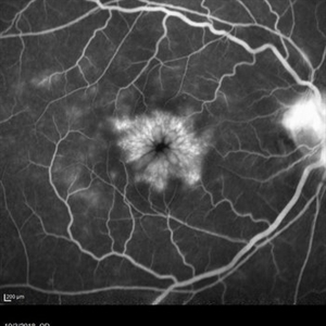 Cystoid Macular Edema Secondary to Panuveitis
Jan 15 2019 by Olivia Rainey
Fluorescein angiogram of a 55-year-old female with cystoid macular edema secondary to uveitis affecting her right eye. Patient was diagnosed with sarcoidosis.
Photographer: Olivia Rainey
Imaging device: Heidelberg Spectralis
Condition/keywords: 30 degrees, cystoid macular edema (CME), fluorescein angiogram (FA), fluorescein leakage, Heidelburg Spectralis, sarcoidosis, uveitis
-
 Multifocal Choroiditis with Panuveitis
Feb 25 2018 by Armando L. Oliver, MD
Multifocal choroiditis with panuveitis. Suppressed on therapy with CellCept, Neoral and Oral Prednisone.
Photographer: Moises Castro
Imaging device: Optos California
Condition/keywords: multifocal choroiditis
-
 Multifocal Choroiditis with Panuveitis
Feb 25 2018 by Armando L. Oliver, MD
Multifocal choroiditis with panuveitis. Suppressed on therapy with CellCept, Neoral and Oral Prednisone.
Photographer: Moises Castro
Imaging device: Optos California
Condition/keywords: multifocal choroiditis
 Loading…