Search results (49 results)
-
---thumb.jpg/image-square;max$300,300.ImageHandler) Multifocal Choroiditis and Panuveitis Syndrome
Multifocal Choroiditis and Panuveitis Syndrome
Feb 26 2013 by Henry J. Kaplan, MD
Multifocal choroiditis, left eye: multiple punched out scar formations in the posterior pole.
Condition/keywords: multifocal choroiditis, panuveitis
-
 Epiretinal Membrane
Oct 11 2012 by Michael P. Kelly, FOPS
This is a patient with idiopathic panuveitis who developed a visually significant epiretinal membrane. Pars plana vitrectomy with membrane peeling was performed to remove the epiretinal proliferation. I recommend magnifying the image to see the exquisite detail centrally.
Photographer: Michael P. Kelly, FOPS Director, Duke Eye Center Labs, Duke Universtiy Hospital
Imaging device: Zeiss 450Plus
Condition/keywords: epiretinal membrane (ERM), panuveitis
-
 Acute Retinal Periphlebitis and Panuveitis OD
Jul 16 2014 by Deepayan Kar
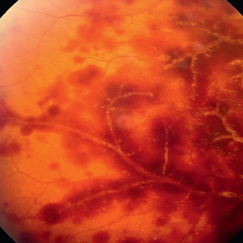
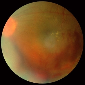
Hemorrhagic retinopathy + frosted retinal angitis OD. Exudative sheathing of the major retinal blood vessels Px 25-year-old woman (VA-OD 20/40/ OS 20/25): recovering from URTI. AC ++ Vit Cells +.
Photographer: Deepayan Kar
Condition/keywords: exudative sheathing, frosted branch angiitis, hemorrhage, retinopathy
-
---thumb.jpg/image-square;max$300,300.ImageHandler) Multifocal Choroiditis & Panuveitis Syndrome
Multifocal Choroiditis & Panuveitis Syndrome
Feb 26 2013 by Henry J. Kaplan, MD
Multifocal choroiditis (MFC) and panuveitis.
Condition/keywords: multifocal choroiditis, panuveitis
-
 Tuberculosis Panuveitis
Feb 25 2013 by Henry J. Kaplan, MD
Peripheral vascular sheathing and multiple choroidiris foci in a patient with tuberculosis panuveitis.
Condition/keywords: panuveitis, tuberculosis
-
---thumb.jpg/image-square;max$300,300.ImageHandler) Multifocal Choroiditis and Panuveitis Syndrome
Multifocal Choroiditis and Panuveitis Syndrome
Feb 26 2013 by Henry J. Kaplan, MD
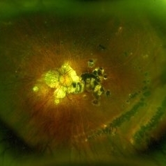
Multifocal choroiditis and panuveitis: left eye. Acute stage: haziness of the media due to vitritis and multiple active yellow and also inactive choroidal lesions are present.
Condition/keywords: multifocal choroiditis
-
---thumb.jpg/image-square;max$300,300.ImageHandler) Multifocal Choroiditis & Panuveitis Syndrome
Multifocal Choroiditis & Panuveitis Syndrome
Feb 26 2013 by Henry J. Kaplan, MD
Multifocal choroiditis, MFC. Lesions look similar to POHS but the patient has vitritis in contrast to the former.
Condition/keywords: multifocal choroiditis, panuveitis
-
---thumb.jpg/image-square;max$300,300.ImageHandler) Multifocal Choroiditis & Panuveitis Syndrome
Multifocal Choroiditis & Panuveitis Syndrome
Feb 26 2013 by Henry J. Kaplan, MD
Multifocal choroiditis, MFC, old scars.
Condition/keywords: multifocal choroiditis, panuveitis
-
 Syphilitic Panuveitis, Left Eye
Oct 8 2012 by Pauline T Merrill, MD, FASRS
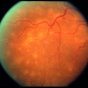
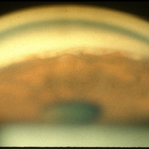
Left fundus photograph of a 23-year-old Hispanic male with decreased vision for 1 month, recent pain/redness both eyes. Found to have panuveitis; also rash on palms & soles. Labs positive for FTA-ABS, and CSR VDRL. Treated with IV penicillin for 14 days.
Photographer: Karen Parque, Illinois Retina Associates, Chicago, IL
Condition/keywords: panuveitis, snowballs, syphilis, vitritis
-
 Syphilitic Panuveitis, Right Eye
Oct 8 2012 by Pauline T Merrill, MD, FASRS
Right fundus photograph of a 23-year-old Hispanic male with decreased vision for 1 month, recent pain/redness both eyes. Found to have panuveitis; also rash on palms & soles. Labs positive for FTA-ABS, and CSR VDRL. Treated with IV penicillin for 14 days.
Photographer: Karen Parque, Illinois Retina Associates, Chicago, IL
Condition/keywords: panuveitis, snowballs, syphilis, vitritis
-
 Sarcoidosis Panuveitis Slide 6
Oct 22 2012 by Ronald C. Gentile, MD
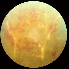
Fundus photo of the peripheral retina of the left eye reveal peri-vascular sheathing and ischemic vasculitis.
Photographer: The New York Eye & Ear Infirmary Department of Medical Imaging
Condition/keywords: sarcoidosis panuveitis, sarcoidosis vasculitis
-
 Sarcoidosis Panuveitis Slide 2
Oct 22 2012 by Ronald C. Gentile, MD
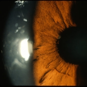
Anterior segment photo of the iris and pupillary margin shows a Koeppe nodule at the 9:30 position. Koeppe nodules consist of inflammatory cell precipitates.
Photographer: The New York Eye & Ear Infirmary Department of Medical Imaging
Condition/keywords: sarcoidosis panuveitis
-
 Occlusive Vasculitis
Mar 9 2013 by Gabriela Lopezcarasa Hernandez, MD
A 36-year-old women with panuveitis and bilateral occlusive vasculitis.
Photographer: Azucena Rios Macula Retina Consultores Mexico
Imaging device: heidelberg Spectralis
Condition/keywords: occlusive vasculitis
-
 Sarcoidosis Panuveitis Slide 5
Oct 22 2012 by Ronald C. Gentile, MD
Fundus photo of the left eye reveal signs of vasculitis with exudate and a vitreous hemorrhage.
Photographer: The New York Eye & Ear Infirmary Department of Medical Imaging
Condition/keywords: sarcoidosis panuveitis, sarcoidosis vasculitis, vitreous hemorrhage
-
---thumb.jpg/image-square;max$300,300.ImageHandler) Multifocal Choroiditis and Panuveitis (MCP) (OS)
Multifocal Choroiditis and Panuveitis (MCP) (OS)
Apr 1 2014 by Min Kim, MD, PhD, MBA, FASRS
Wide field fundus photograph of a 61-year-old female with decreased vision in both eyes due to multifocal choroiditis and panuveitis.
Condition/keywords: multifocal choroiditis, panuveitis
-
 Sarcoidosis Panuveitis Slide 3
Oct 22 2012 by Ronald C. Gentile, MD
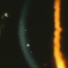
Gonioscopic photograph reveals peripheral anterior synechiae with granuloma involving the iris root and Bussaca nodules on the iris surface consistent with granulomatous uveitis.
Photographer: The New York Eye & Ear Infirmary Department of Medical Imaging
Condition/keywords: sarcoid granuloma, sarcoidosis panuveitis
-
 Multifocal Choroiditis and Panuveitis- Schlaegel lines
Nov 16 2021 by Manuel Ángel Alcántara Delgado, MD
Optomap ultra-widefield retinal imaging of an 52-year-old woman showed multiple punched-out chorioretinal lesions and 2 rows of peripheral curvilinear pigmented chorioretinal streaks (Schlaegel lines).
Photographer: Manuel Ángel Alcántara Delgado. Conde de Valenciana.
Condition/keywords: multifocal choroiditis, myopia, retina, uveitis
-
 Sarcoidosis Panuveitis Slide 4
Oct 22 2012 by Ronald C. Gentile, MD
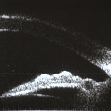
High frequency ultrasound biomicroscopy of the anterior chamber and angle images a granuloma involving the iris root and Bussaca nodules on the iris surface consistent with granulomatous uveitis.
Photographer: The New York Eye & Ear Infirmary Department of Medical Imaging
Condition/keywords: sarcoid granuloma, sarcoidosis panuveitis
-
 Sarcoidosis Panuveitis Slide 1
Oct 22 2012 by Ronald C. Gentile, MD
50-year-old African American women presented with decreasing vision in both eyes and floaters in the left eye. She had a history of asthma and sub-cutaneous skin nodules. Anterior segment examination reveal keratic precipitate on the corneal endothelium.
Photographer: The New York Eye & Ear Infirmary Department of Medical Imaging
Condition/keywords: sarcoidosis panuveitis
-
 Sarcoidosis Panuveitis Slide 7
Oct 22 2012 by Ronald C. Gentile, MD
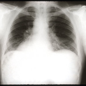
Chest x-ray revealed bihilar lymphadenopathy.
Photographer: The New York Eye & Ear Infirmary Department of Radiology
Condition/keywords: sarcoidosis panuveitis
-
 Vogt-Koyanagi-Harada Disease
Feb 13 2019 by Deepak Bhojwani, MS
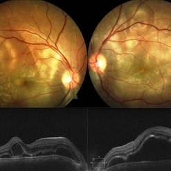
A 18-year-old girl came with complaints of acute onset of diminution of vision associated with dull boring pain and headache. She gave a history of flu like symptoms with fever headache and malaise few days before onset of ocular symptoms. Fundus photographs shows classic picture of acute stage of VKH with multiple large serous RPE detachments and inflamed choroid. OCT shows hallmark features of VKF viz. multiple serous RPEDs with hyperreflective dots, subretinal septa and thick choroid.
Photographer: DR DEEPAK BHOJWANI
Imaging device: Zeiss Visucam 500
Condition/keywords: bilateral serous detachment, panuveitis, Vogt-Koyanagi-Harada
-
 Sarcoidosis Panuveitis Slide 8
Oct 22 2012 by Ronald C. Gentile, MD
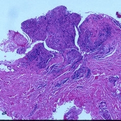
Biopsy of one of the sub-cutaneous nodules revealed non-caseating granulomas consistent with sarcoidosis.
Photographer: The New York Eye & Ear Infirmary Department of Pathology and Laboratory Medicine
Condition/keywords: sarcoidosis panuveitis
-
---thumb.jpg/image-square;max$300,300.ImageHandler) Multifocal Choroiditis and Panuveitis (MCP)(1)
Multifocal Choroiditis and Panuveitis (MCP)(1)
Apr 1 2014 by Min Kim, MD, PhD, MBA, FASRS
Wide field fundus photograph of a 61-year-old female with decreased vision in both eyes due to multifocal choroiditis and panuveitis.
Condition/keywords: multifocal choroiditis, panuveitis
-
 Multifocal Choroiditis and Panuveitis (MCP)
Apr 1 2014 by Min Kim, MD, PhD, MBA, FASRS
Wide field fundus photograph of a 61-year-old female with decreased vision in both eyes due to multifocal choroiditis and panuveitis.
Photographer: Young Duk Bae, Yonsei University, Gangnam Severance Hospital
Imaging device: Optos
Condition/keywords: multifocal choroiditis, panuveitis
-
---thumb.jpg/image-square;max$300,300.ImageHandler) Multifocal Choroiditis and Panuveitis (MCP)(OD)
Multifocal Choroiditis and Panuveitis (MCP)(OD)
Apr 1 2014 by Min Kim, MD, PhD, MBA, FASRS
Wide field fundus photograph of a 61-year-old female with decreased vision in both eyes due to multifocal choroiditis and panuveitis.
Condition/keywords: multifocal choroiditis, panuveitis
 Loading…