Search results (49 results)
-
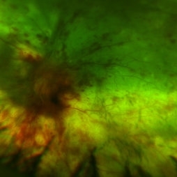
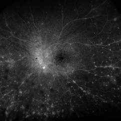
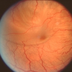
 Epiretinal Membrane
Oct 11 2012 by Michael P. Kelly, FOPS
This is a patient with idiopathic panuveitis who developed a visually significant epiretinal membrane. Pars plana vitrectomy with membrane peeling was performed to remove the epiretinal proliferation. I recommend magnifying the image to see the exquisite detail centrally.
Photographer: Michael P. Kelly, FOPS Director, Duke Eye Center Labs, Duke Universtiy Hospital
Imaging device: Zeiss 450Plus
Condition/keywords: epiretinal membrane (ERM), panuveitis
-
---thumb.jpg/image-square;max$300,300.ImageHandler) Multifocal Choroiditis & Panuveitis Syndrome
Multifocal Choroiditis & Panuveitis Syndrome
Feb 26 2013 by Henry J. Kaplan, MD
Multifocal choroiditis (MFC) and panuveitis.
Condition/keywords: multifocal choroiditis, panuveitis
-
---thumb.jpg/image-square;max$300,300.ImageHandler) Multifocal Choroiditis & Panuveitis Syndrome
Multifocal Choroiditis & Panuveitis Syndrome
Feb 26 2013 by Henry J. Kaplan, MD
Multifocal choroiditis, MFC. Lesions look similar to POHS but the patient has vitritis in contrast to the former.
Condition/keywords: multifocal choroiditis, panuveitis
-
---thumb.jpg/image-square;max$300,300.ImageHandler) Multifocal Choroiditis & Panuveitis Syndrome
Multifocal Choroiditis & Panuveitis Syndrome
Feb 26 2013 by Henry J. Kaplan, MD
Multifocal choroiditis, MFC, old scars.
Condition/keywords: multifocal choroiditis, panuveitis
-
 Multifocal Choroiditis and Panuveitis (MCP)
Apr 1 2014 by Min Kim, MD, PhD, MBA, FASRS
Wide field fundus photograph of a 61-year-old female with decreased vision in both eyes due to multifocal choroiditis and panuveitis.
Photographer: Young Duk Bae, Yonsei University, Gangnam Severance Hospital
Imaging device: Optos
Condition/keywords: multifocal choroiditis, panuveitis
-
---thumb.jpg/image-square;max$300,300.ImageHandler) Multifocal Choroiditis and Panuveitis (MCP) (OS)
Multifocal Choroiditis and Panuveitis (MCP) (OS)
Apr 1 2014 by Min Kim, MD, PhD, MBA, FASRS
Wide field fundus photograph of a 61-year-old female with decreased vision in both eyes due to multifocal choroiditis and panuveitis.
Condition/keywords: multifocal choroiditis, panuveitis
-
---thumb.jpg/image-square;max$300,300.ImageHandler) Multifocal Choroiditis and Panuveitis (MCP)(1)
Multifocal Choroiditis and Panuveitis (MCP)(1)
Apr 1 2014 by Min Kim, MD, PhD, MBA, FASRS
Wide field fundus photograph of a 61-year-old female with decreased vision in both eyes due to multifocal choroiditis and panuveitis.
Condition/keywords: multifocal choroiditis, panuveitis
-
---thumb.jpg/image-square;max$300,300.ImageHandler) Multifocal Choroiditis and Panuveitis (MCP)(OD)
Multifocal Choroiditis and Panuveitis (MCP)(OD)
Apr 1 2014 by Min Kim, MD, PhD, MBA, FASRS
Wide field fundus photograph of a 61-year-old female with decreased vision in both eyes due to multifocal choroiditis and panuveitis.
Condition/keywords: multifocal choroiditis, panuveitis
-
---thumb.jpg/image-square;max$300,300.ImageHandler) Multifocal Choroiditis and Panuveitis Syndrome
Multifocal Choroiditis and Panuveitis Syndrome
Feb 26 2013 by Henry J. Kaplan, MD
Multifocal choroiditis, left eye: multiple punched out scar formations in the posterior pole.
Condition/keywords: multifocal choroiditis, panuveitis
-
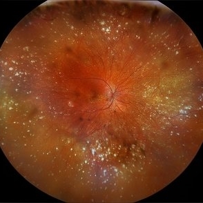
 Panuveitis
Jul 12 2024 by Korey Starkey
Ultra widefield Optos FA of 59 year old female presents with panuveitis in both eyes. Patients vision was VA OS: Dcc20/60-2 at time of visit.
Photographer: Korey Starkey
Imaging device: Optos
Condition/keywords: FLUORESCEIN ANGIOGRAPHY, hyperfluorescence, Optos, Panuveitis, ultra-wide field imaging, Uveitis
-
 Panuveitis
Apr 2 2024 by Zach Seim
Optos Ultra-widefield photo OS of a 59 year old female with Panuveitis OU.
Photographer: Zach Seim
Imaging device: Optos California
Condition/keywords: Optos, OPTOS CALIFORNIA, panuveitis, ULTRA WIDE FIELD, ultra-wide field imaging
-
 Panuveits / Disseminated Chorioretinitis
Nov 24 2015 by Matt Poe, COA
This is of a 42-year-old woman that presented to our office with visual acuity of NLP. Labs show that patient is positive for cytomegalovirus. Labs with increased IgG titer for CMV, but normal IgM. She has been diagnosed with Panuveitis, Disseminated chorioretinitis, and serous retinal detachment.
Photographer: Matt Poe, COA. Northwest Arkansas Retina Associates, Springdale, AR.
Condition/keywords: panuveitis, serous retinal detachment
-
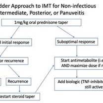 Posterior Manifestations of Sarcoidosis – Management of noninfectious intermediate, posterior, or panuveitis
Mar 29 2023 by Joshua Friedman
Management of noninfectious intermediate, posterior, or panuveitis.
Condition/keywords: panuveitis, sarcoidosis
-
 Resolved Exudative RD in Vogt-Koyanagi-Harada Syndrome
Mar 27 2019 by Gary R. Cook, MD, FACS
Right eye of the same patient following resolution of the exudative retinal detachment OD.
Condition/keywords: exudative detachment, panuveitis, Vogt-Koyanagi-Harada
-
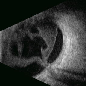 Stag Horn
Apr 8 2025 by Gustavo Uriel Fonseca Aguirre
B-mode ultrasound of a young male patient with bilateral panuveitis (currently under investigation) reveals intense vitritis with islands of preserved vitreous and partial posterior hyaloid detachment, creating a characteristic "stag horn" appearance.
Photographer: Gustavo U. Fonseca Aguirre, Hospital Conde de Valenciana, Ciudad de México
Condition/keywords: Panuveitis
-
 Syphilitic Panuveitis, Left Eye
Oct 8 2012 by Pauline T Merrill, MD, FASRS
Left fundus photograph of a 23-year-old Hispanic male with decreased vision for 1 month, recent pain/redness both eyes. Found to have panuveitis; also rash on palms & soles. Labs positive for FTA-ABS, and CSR VDRL. Treated with IV penicillin for 14 days.
Photographer: Karen Parque, Illinois Retina Associates, Chicago, IL
Condition/keywords: panuveitis, snowballs, syphilis, vitritis
-
 Syphilitic Panuveitis, Right Eye
Oct 8 2012 by Pauline T Merrill, MD, FASRS
Right fundus photograph of a 23-year-old Hispanic male with decreased vision for 1 month, recent pain/redness both eyes. Found to have panuveitis; also rash on palms & soles. Labs positive for FTA-ABS, and CSR VDRL. Treated with IV penicillin for 14 days.
Photographer: Karen Parque, Illinois Retina Associates, Chicago, IL
Condition/keywords: panuveitis, snowballs, syphilis, vitritis
-
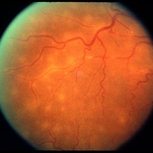 Tuberculosis Panuveitis
Feb 25 2013 by Henry J. Kaplan, MD
Peripheral vascular sheathing and multiple choroidiris foci in a patient with tuberculosis panuveitis.
Condition/keywords: panuveitis, tuberculosis
-
 VKH - Uveitic stage
Jun 23 2023 by Sergio Emilio Sifuentes Renteria, MD
Fundus photograph of a young female with VKH in uveitic stage
Photographer: Sergio Emilio Sifuentes Rentería - Foundation Hospital Nuestra Señora de La Luz
Condition/keywords: choroiditis, panuveitis, serous retinal detachment, VKH, Vogt-Koyanagi-Harada
-
 VKH Syndrome
Jun 12 2025 by Virginia Gebhart
Fluorescein angiogram of 22 year old male with VKH syndrome. Significant cell in AC and vitreous, multiple punched-out CR scars in periphery, mild vascular leakage. Pt referred to rheumatology for immunomodulatory treatment.
Photographer: Virginia Gebhart, Retina Consultants of Carolina
Imaging device: Optos California
Condition/keywords: FA, fluorescein angiogram (FA), multifocal choroiditis, panuveitis, VKH, Vogt-Koyanagi-Harada
-
 VKH Syndrome
Jun 12 2025 by Virginia Gebhart
22 year old male with VKH Syndrome. Pt has been experiencing severe headaches, distorted vision, hearing loss, weakness, and a large white patch of hair. Significant cell in AC and vitreous, multiple punched-out CR scars in periphery. Referred to rheumatology for possible immunomodulatory treatment
Photographer: Virginia Gebhart, Retina Consultants of Carolina
Imaging device: Optos California
Condition/keywords: montage, multifocal choroiditis, panuveitis, Vogt-Koyanagi-Harada
-
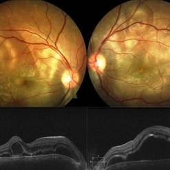 Vogt-Koyanagi-Harada Disease
Feb 13 2019 by Deepak Bhojwani, MS
A 18-year-old girl came with complaints of acute onset of diminution of vision associated with dull boring pain and headache. She gave a history of flu like symptoms with fever headache and malaise few days before onset of ocular symptoms. Fundus photographs shows classic picture of acute stage of VKH with multiple large serous RPE detachments and inflamed choroid. OCT shows hallmark features of VKF viz. multiple serous RPEDs with hyperreflective dots, subretinal septa and thick choroid.
Photographer: DR DEEPAK BHOJWANI
Imaging device: Zeiss Visucam 500
Condition/keywords: bilateral serous detachment, panuveitis, Vogt-Koyanagi-Harada
-
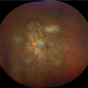 Vogt-Koyanagi-Harada Disease
Apr 24 2022 by Aniruddha K Agarwal, MD
A 38-year-old woman of Asian descent with no ophthalmological or systemic history presented to the emergency eye clinic with a 1-week complaint of headache and bilateral vision loss. Funduscopy revealed bilateral serous neurosensory detachments. The presence of lymphocytosis in cerebrospinal fluid and mild acute sensorineural hearing loss confirmed the diagnosis of uveomeningoencephalitic syndrome (Vogt-Koyanagi-Harada disease).
Photographer: Mercedes SERRADOR, MD, PhD and Beatriz VENTAS, MD
Imaging device: Zeiss Clarus fundus camera
Condition/keywords: IUSG, panuveitis, Vogt-Koyanagi-Harada
-
 Vogt-Koyanagi-Harada Syndrome
Mar 27 2019 by Gary R. Cook, MD, FACS
Exudative retinal detachment secondary to Vogt-Koyanagi-Harada syndrome OD
Condition/keywords: exudative detachment, panuveitis, Vogt-Koyanagi-Harada
-
 Vogt-Koyanagi-Harada Syndrome
Mar 27 2019 by Gary R. Cook, MD, FACS
View along superior arcade of exudative retinal detachment secondary to Vogt-Koyanagi-Harada Syndrome OD.
Condition/keywords: exudative detachment, panuveitis, Vogt-Koyanagi-Harada
 Loading…