Search results (556 results)
-
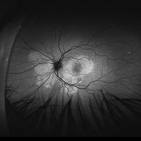
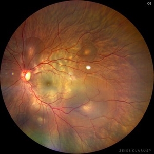
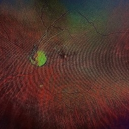
 Ocular Melanocytosis w/Treated Melanoma
Jan 27 2025 by Virginia Gebhart
74 year female with ocular melanocytosis. Stable, regressed treated tumor s/p brachytherapy (2020) and deeply pigmented fundus OS. Limited VA due to radiation neuropathy. BCVA 20/150 (ecc)
Photographer: Virginia Gebhart, Retina Consultants of Carolina
Imaging device: Optos California
Condition/keywords: brachytherapy, melanoma, melanosis, ocular melanocytosis
-
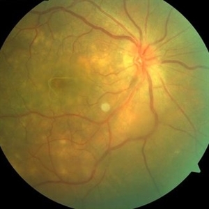
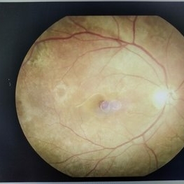
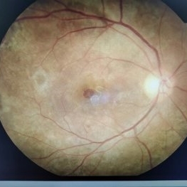
 Hamartoma of the Retina and Retinal Pigment Epithelium
Jan 5 2025 by César Adrián Gómez Valdivia, MD
Hamartoma of the retina and retinal pigment epithelium found in a 10 year-old male patient with type 2 neurofibromatosis history. Overlaying fibrous proliferation can be appreciated. Findings were unilateral.
Photographer: @eyemissu2
Imaging device: TOPCON TRC-50DX
Condition/keywords: hamartoma, retinal pigment epithelium (RPE) hamartoma
-
 Acute Syphilitic Posterior Placoid Chorioretinitis
Oct 20 2024 by César Adrián Gómez Valdivia, MD
Fundus autofluorescence image of an acute syphilitic posterior placoid chorioretinitis found in a HIV positive 28 YO male patient with suspected neurosyphilis. A beautiful butterfly autofluorescence pattern can be appreciated.
Photographer: @eyemissu2
Imaging device: California ICG OPTOS
Condition/keywords: acute syphilitic posterior placoid chorioretinitis
-
 Acute Syphilitic Posterior Placoid Chorioretinitis
Oct 16 2024 by César Adrián Gómez Valdivia, MD
Fundus autofluorescence image of an acute syphilitic posterior placoid chorioretinitis found in a HIV positive 28 YO male patient with suspected neurosyphilis. A beautiful butterfly autofluorescence pattern can be appreciated.
Photographer: @eyemissu2
Imaging device: California ICG OPTOS
Condition/keywords: acute syphilitic posterior placoid chorioretinitis, chorioretinitis, syphilis
-
 Worm Eye (DUSN)
Sep 11 2024 by lucas valadao soares, md
Fundus photograph of an 6-year-old boy with a nematode in the left eye.
Photographer: Lucas Valadao, Brasil
Imaging device: Canon CX-1
Condition/keywords: diffuse unilateral subacute neuroretinitis (DUSN)
-
 DUSN
DUSN
Aug 4 2024 by Sharat Shivaramaiah Hegde, MS OPHTHALMOLOGY
41 year old with sudden loss of vision noted to have large worm in submacular space with neuosensory detachment with multiple outer retinal crops and disc edema.
Photographer: Sharat Hegde , Prasad Netralaya, Udupi
Imaging device: Visucam 500
Condition/keywords: diffuse unilateral subacute neuroretinitis (DUSN), nematode
-
 Focal Chorioretinitis
Jul 11 2024 by Virginia Gebhart
67 year old female with punched-out CR scars. Hx of laser 3x for apparent peripapillary CNV. ESR, CRP, toxo, IgG/IgM all "normal." Bartonella, quant gold, and FTA-ABS ordered given possibility of neuroretinitis. Vision CF
Photographer: Virginia Gebhart
Imaging device: Optos California
Condition/keywords: FA, fluorescein angiogram (FA), FLUORESCEIN ANGIOGRAPHY, focal chorioretinitis, optic neuritis
-
 Central Serous Chorioretinopathy in Pregnancy (OD)
Apr 28 2024 by Vishal Agrawal, MD, FRCS,FACS,FASRS
30-year female with sudden loss of vision came for examination. She was in her first trimester of pregnancy. Examination revealed bilateral bullous NSD with subretinal fibrin s/o CSR.
Photographer: Dr Ayushi
Imaging device: Clarus 700
Condition/keywords: Central Serous Chorioretinopathy (CSR), neurosensory detachment of retina, pregnancy
-
 Central Serous Chorioretinopathy in Pregnancy (OS)
Apr 28 2024 by Vishal Agrawal, MD, FRCS,FACS,FASRS
30-year female with sudden loss of vision came for examination. She was in her first trimester of pregnancy. Examination revealed bilateral bullous NSD with subretinal fibrin s/o CSR.
Photographer: Dr Ayushi
Imaging device: Clarus 700
Condition/keywords: Central Serous Chorioretinopathy (CSR), neurosensory detachment of retina, pregnancy
-
 Acute Macular Neuroretinopathy
Mar 25 2024 by Daniel Davis, OCT-C
18 yo female presenting with hazy vison for 2-3 weeks. VA OD: sc20/20 VA OS: sc20/20 Infrared imaging showed dark gray, petalloid, perifoveal lesions and OCT shows focal signal reduction of the Inner Segment / Outer Segment junction. Elects to observe.
Photographer: Daniel Davis, OCT-C, The Retina Institute, St. Louis
Imaging device: Optos California SWL
Condition/keywords: acute macular neuroretinopathy
-
 NF1
NF1
Mar 19 2024 by Majda Hadziahmetovic, MD
Near-infrared reflectance imaging shows the presence of choroidal hyperreflective nodules in a patient diagnosed with NF1.
Condition/keywords: Neurofibromatosis 1
-
 Venous Loop
Feb 20 2024 by Soobien Lee
A 77-year-old male with a history of bilateral optic neuropathy from bilateral optic nerve sheath meningiomas S/P radiation/proton-beam therapies. Presented with radiation retinopathy OS and a known venous loop OS.
Photographer: Gavin Bragdon, Elman Retina Group
Imaging device: Optos Ultra-Widefield Fluorescein Angiography
Condition/keywords: fluorescein angiogram (FA), Optos, radiation retinopathy, retinal vascular disease, venous loop
-
 Venous Loop
Feb 20 2024 by Soobien Lee
A 77-year-old male with a history of bilateral optic neuropathy from bilateral optic nerve sheath meningiomas S/P radiation/proton-beam therapies. Presented with radiation retinopathy OS and a known venous loop OS.
Photographer: Gavin Bragdon, Elman Retina Group
Imaging device: Optos Ultra-Widefield Imaging
Condition/keywords: Optos, OPTOS CALIFORNIA, radiation retinopathy, retinal vascular disease, venous loop
-
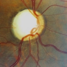
 Morning Glory Disc Anomaly
Feb 12 2024 by NIDHI PANWAR, MD FNB FICO
Fundus photograph of 43 year old male, hypertensive on medication, came for routine check up, and has been diagnosed to have poor vision left eye since childhood, denies any history of trauma. Vision left eye 6/18, Anterior segment normal, Fundus left eye shows excavated ,funnel-shaped optic nerve head, with central tuft of glial tissue obscuring the cup . The retinal vessels were seen emanating from the edge of disc in radial manner. In addition, the sectoral nasal retina shows localized area of hyperpigmented bony spicules like lesions. However, no history of nyctalopia or any other neurological disorder could be obtained.
Photographer: Nidhi Panwar, NMC Royal hospital, Sharjah , UAE
Imaging device: OPTOMAP
Condition/keywords: Morning Glory Anomaly, optic disc excavation
-
 Joubert Syndrome
Jan 20 2024 by Francisco Fraga Santini Canto
Fundus autofluorescence of a 57-year-old woman with macular distrophy in both eyes. Patient also had neurological findings. Genetic test showed a pathogenic mutation of gene AHI1 confirming Joubert Syndrome.
Photographer: Leonardo Hideki Nomachi Naito
Imaging device: Silverstone - Ultra-widefield
Condition/keywords: AH1 mutation, autossomalrecessive, ciliopathies, genetcis, joubert syndrome, macular distrophy
-
 Left Inferior Quadrantopia - Microperimetry
Jan 17 2024 by Francisco Fraga Santini Canto
Microperimetry of a 63-year-old male with sequelae of ischemic stroke in the right parietal lobe years ago.
Photographer: Leonardo Hideki Nomachi Naito
Imaging device: Navis-EX Microperimeter
Condition/keywords: ischemic stroke, microperimetry, neuro, neuro-ophtalmology, visual field defect
-
 Left Inferior Quadrantopia - Microperimetry
Jan 17 2024 by Francisco Fraga Santini Canto
Microperimetry of a 63-year-old male with sequelae of ischemic stroke in the right parietal lobe years ago. In the right eye, the patient also had a central scotoma secondary to silicone oil toxicity.
Photographer: Leonardo Hideki Nomachi Naito
Imaging device: Navis-EX Microperimeter
Condition/keywords: ischemic stroke, microperimeter, neuro-ophtalmolgy, silicone oil toxicity, visual field defect
-
 Central Serous Chorioretinopathy (CSR)
Sep 21 2023 by Ben Serar
Fundus photograph showing increased cup-disc ratio with nasalisation of vessels , with thinning of Neuroretinal rim and bayonetting of vessels in a case of Glaucomatous Optic Atrophy (GOA) Fundus photograph of LE showing serous macular detachment in a case of Central Serous Chorioretinopathy (CSR).
Condition/keywords: Central Serous Chorioretinopathy (CSR)
-
 Glaucomatous Optic Atrophy (GOA)
Sep 21 2023 by Ben Serar
Fundus photograph showing increased cup-disc ratio with nasalisation of vessels , with thinning of Neuroretinal rim and bayonetting of vessels in a case of Glaucomatous Optic Atrophy (GOA).
Condition/keywords: Glaucomatous Optic Atrophy (GOA)
-
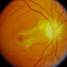 Neuroretinitis
Sep 21 2023 by Ben Serar
Fundus photograph of the RE showing Disc edema with blurring of disc margins with pallor, with exudates at the macula arranged in a star-shaped pattern, in a case of Neuroretinitis.
Condition/keywords: macular star, neuroretinitis
-
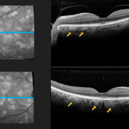
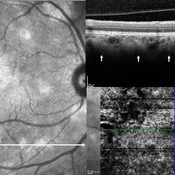 Choroidal Nodules in Neurofibromatosis
Sep 6 2023 by Maria Filipa Madeira
Macular near-infrared reflectance (NIR) imaging, optical coherence tomography (OCT) B-scan and OCT angiography (OCTA) of a 54-year-old woman with neurofibromatosis type 1. Choroidal abnormalities were asymptomatic and not visible on funduscopic exam, but had a striking appearance on retinal imaging. B-scan (horizontal arrow) showed hyperreflective nodules in the deeper choroid (vertical arrows) underlying the multiple hyperreflective patches on NIR, in correlation with hyperflow areas of the deep choroidal plexus in OCTA.
Photographer: Maria Filipa Madeira, Centro Hospitalar de Lisboa Ocidental, Hospital de Egas Moniz
Imaging device: Heidelberg Spectralis
Condition/keywords: choroid, neurofibromatosis
-
 Diffuse Unilateral Subacute Neuroretinitis
May 20 2023 by DITSHA DATTA
Fundus photograph of a 16 year old female with diffuse unilateral subacute neuroretinitis
Photographer: Ditsha Datta,GSVM Medical College, India
Imaging device: Smart phone fundoscopy
Condition/keywords: diffuse unilateral subacute neuroretinitis (DUSN)
-
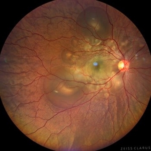
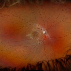
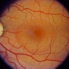
 Optic Nerve Melanocytoma
Apr 3 2023 by Gustavo Aguirre Suarez
Fundus photograph of a 36-year-old female with a lesion dependent on the optic nerve head with subretinal extension, elevated, about 1.5 disc diameters, dark brown to black in color, involving more than three quarters of the neuroretinal ring towards the inferonasal area.
Photographer: Dr. Gustavo Aguirre-Suarez
Imaging device: Zeiss Visucam 500
Condition/keywords: melanocytic lesion, Melanocytoma
 Loading…