Search results (556 results)
-
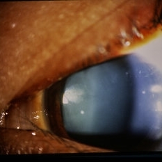
 Left Inferior Quadrantopia - Microperimetry
Jan 17 2024 by Francisco Fraga Santini Canto
Microperimetry of a 63-year-old male with sequelae of ischemic stroke in the right parietal lobe years ago.
Photographer: Leonardo Hideki Nomachi Naito
Imaging device: Navis-EX Microperimeter
Condition/keywords: ischemic stroke, microperimetry, neuro, neuro-ophtalmology, visual field defect
-
 Capillary Hemangioma
Mar 27 2019 by Gary R. Cook, MD, FACS
White male with a retinal capillary hemangioma OD secondary to neurofibromatosis.
Condition/keywords: neurofibromatosis, retinal hemangioma
-
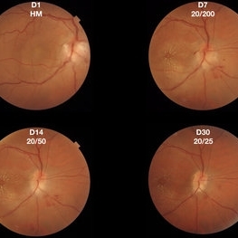 Cat-Scratch Neuroretinitis
Nov 1 2017 by FELIPE PEREIRA
40-year-old patient presented with sudden and painless visual acuity loss for 1 day. His initial visual acuity was hand motion. There was positive epidemiology for cat-scratch disease and other serologies tests were negative. Treatment was initiated with 200 mg/day of Doxycycline for 21 days plus oral prednisone 1mg/kg tapered in 4 weeks. The patient presented a favorable evolution with reduction of the peripapillary granuloma and improvement of visual acuity to 20/25.
Photographer: Felipe Pereira
Imaging device: Triton, Topcon
Condition/keywords: neuroretinitis, ocular bartonellosis
-
 EDI-Optic Neuritis/Neuroretinitis
Jul 15 2013 by Jason S. Calhoun
Patient with some loss of vision in his left eye last week, was seen by eye MD and referred for eval. Patient also complained of pain in left eye with eye movement. VA was 20/400 in the left eye. Fundus photo and HD-OCT imaging show optic nerve swelling and fluid underneath the retina. A neuro-ophthalmologist will be consulted for further evaluation.
Photographer: Jason S. Calhoun, Department of Ophthalmology, Mayo Clinic Jacksonville, Florida
Imaging device: ZEISS OCT CIRRUS
Condition/keywords: neuroretinitis, optic neuritis
-
 EDI-Optic Neuritis/Neuroretinitis
Jul 15 2013 by Jason S. Calhoun
Patient with some loss of vision in his left eye and was seen for an evaluation. Patient also complained of pain in left eye with eye movement. VA was 20/400 in the left eye. Fundus photo and HD-OCT imaging show optic nerve swelling and fluid underneath the retina. A neuro-ophthalmologist will be consulted for further evaluation.
Photographer: Jason S. Calhoun, Department of Ophthalmology, Mayo Clinic Jacksonville, Florida
Imaging device: ZEISS OCT CIRRUS
Condition/keywords: neuroretinitis, optic neuritis
-
 Facial Neurofibroma
Feb 4 2015 by H. Michael Lambert, MD
Extensive facial neurofibroma in patient with neurofibromatosis.
Condition/keywords: neurofibromatosis
-
 Facial Neurofibroma
Feb 4 2015 by H. Michael Lambert, MD
Extensie facial neurofibroma in patient with neurofibromatosis.
Condition/keywords: neurofibromatosis
-
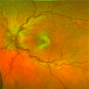 Idiopathic Neuroretinitis
May 6 2020 by David L Kilpatrick, MD
Fundus photo of a 25-year-old white female with idiopathic neuroretinitis.
Photographer: RCA
Imaging device: Optos
Condition/keywords: neuroretinitis
-
 Neurocysticercosis
Sep 10 2020 by Anamika Dwivedi
MRI images of a 22-year-old male with h/o headache for 1 month. MRI brain axial scan showing multiple tiny cystic lesions scattered all over the cerebral parenchyma. Post gadolinium contrast coronal scan showing multiple cystic lesion scattered in cerebral parenchyma, few of the lesion showing ring enhancement.
Condition/keywords: neurocysticercosis
-
 Neurofibroma Pathology Slide
Feb 4 2015 by H. Michael Lambert, MD
Pathologic slide of neurofibroma.
Condition/keywords: neurofibromatosis, pathology
-
 Neurofibroma Pathology Slide
Feb 4 2015 by H. Michael Lambert, MD
Pathologic slide of neurofibroma.
Condition/keywords: neurofibromatosis
-
 Neurofibroma Pathology Slide
Feb 4 2015 by H. Michael Lambert, MD
Pathologic slide of neurofibroma.
Condition/keywords: neurofibromatosis, pathology
-
 Neurofibroma Upper Eye Lid
Feb 4 2015 by H. Michael Lambert, MD
Neurofibroma as seen in upper eye lid of patient with neurofibromatosis.
Condition/keywords: neurofibromatosis
-
 Neurofibromas Under Skin
Feb 4 2015 by H. Michael Lambert, MD
Glandular neurofibromas seen under skin in patient with neurofibromatosis.
Condition/keywords: neurofibromatosis
-
 Neurofibromatosis 2
Oct 9 2012 by Audina M. Berrocal, MD FASRS
Typical vertical membranes seen in cases of NF2
Photographer: BPEI
Condition/keywords: neurofibromatosis
-
 Neurofibromatosis II with RPE Hamartoma
Aug 14 2015 by David Callanan, MD
Neurofibromatosis II with RPE hamartoma
Condition/keywords: hamartoma, neurofibromatosis, retinal pigment epithelium
-
 Neurofibromatosis II with RPE Hamartoma
Aug 14 2015 by David Callanan, MD
Neurofibromatosis II with RPE hamartoma
Condition/keywords: hamartoma, neurofibromatosis, retinal pigment epithelium
-
 Neurofibromatosis II with RPE Hamartoma
Aug 14 2015 by David Callanan, MD
Neurofibromatosis II with RPE hamartoma.
Condition/keywords: hamartoma, neurofibromatosis, retinal pigment epithelium
-
 Neurofibromatosis II with RPE Hamartoma
Aug 14 2015 by David Callanan, MD
Neurofibromatosis II with RPE hamartoma.
Condition/keywords: hamartoma, neurofibromatosis, retinal pigment epithelium
-
 Neurofibromatosis II with RPE Hamartoma
Aug 14 2015 by David Callanan, MD
Neurofibromatosis II with RPE hamartoma.
Condition/keywords: hamartoma, neurofibromatosis, retinal pigment epithelium
-
 Neurofibromatosis II with RPE Hamartoma
Aug 14 2015 by David Callanan, MD
Neurofibromatosis II with RPE hamartoma.
Condition/keywords: hamartoma, neurofibromatosis, retinal pigment epithelium
-
 Neurofibromatosis II with RPE Hamartoma
Aug 14 2015 by David Callanan, MD
Neurofibromatosis II with RPE hamartoma.
Condition/keywords: hamartoma, neurofibromatosis, retinal pigment epithelium
-
 Neurofibromatosis II with RPE Hamartoma
Aug 14 2015 by David Callanan, MD
Neurofibromatosis II with RPE hamartoma.
Condition/keywords: hamartoma, neurofibromatosis, retinal pigment epithelium
-
 Neurofibromatosis II with RPE Hamartoma
Aug 14 2015 by David Callanan, MD
Neurofibromatosis II with RPE hamartoma.
Condition/keywords: hamartoma, neurofibromatosis, retinal pigment epithelium
-
 Neurofibromatosis II with RPE Hamartoma
Aug 14 2015 by David Callanan, MD
Neurofibromatosis II with RPE hamartoma.
Condition/keywords: hamartoma, neurofibromatosis, retinal pigment epithelium
 Loading…