Search results (556 results)
-
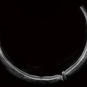 Wide-Field-OCT-montage
Jan 8 2018 by Netan Choudhry, MD, FRCS(C) FASRS
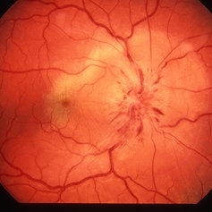
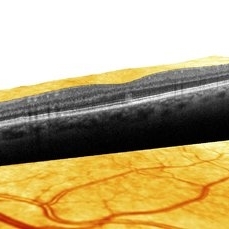
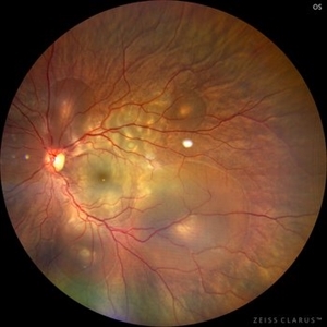
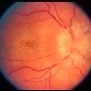
This is an SD-OCT montage image of a 55 year old male with optic neuropathy representing a wide-field OCT spanning 130 degrees.
Photographer: John Golding, Vitreous Retina Macula Specialists of Toronto
Imaging device: Heidelberg Spectralis OCT system
Condition/keywords: wide angle imaging
-
 Venous Loop
Feb 20 2024 by Soobien Lee
A 77-year-old male with a history of bilateral optic neuropathy from bilateral optic nerve sheath meningiomas S/P radiation/proton-beam therapies. Presented with radiation retinopathy OS and a known venous loop OS.
Photographer: Gavin Bragdon, Elman Retina Group
Imaging device: Optos Ultra-Widefield Imaging
Condition/keywords: Optos, OPTOS CALIFORNIA, radiation retinopathy, retinal vascular disease, venous loop
-
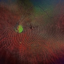
 Venous Loop
Feb 20 2024 by Soobien Lee
A 77-year-old male with a history of bilateral optic neuropathy from bilateral optic nerve sheath meningiomas S/P radiation/proton-beam therapies. Presented with radiation retinopathy OS and a known venous loop OS.
Photographer: Gavin Bragdon, Elman Retina Group
Imaging device: Optos Ultra-Widefield Fluorescein Angiography
Condition/keywords: fluorescein angiogram (FA), Optos, radiation retinopathy, retinal vascular disease, venous loop
-
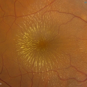 Cat Scratch
Feb 15 2017 by Hua Gao, MD, PhD, FASRS
A female patient of 57-year-old presented with neuroretinitis due to cat-scratch disease with positive Bartonella henselae antibodies. Two weeks after symptom onset she developed exudates in a "macular star" pattern.
Condition/keywords: cat scratch retinitis
-
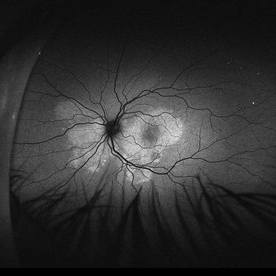
 Acute Macular Neuroretinopathy
Dec 11 2019 by Lauren Whaley
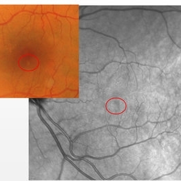
34-year-old female patient presented with changes in vision after recent upper respiratory infection. Referring doctor originally thought it was a blood pressure issue. She noticed a "C" shape in her vision. Infrared image was captured showing exactly what patient was describing! Doctor confirmed with this image that it was AMN.
Photographer: Lauren R. Whaley, COA
Imaging device: Heidelberg Spectralis
Condition/keywords: 30 degrees, acute macular neuroretinopathy, Heidelburg Spectralis, left eye, macula, near infrared autofluorescence (NIRAF)
-
 Acute Syphilitic Posterior Placoid Chorioretinitis
Oct 16 2024 by César Adrián Gómez Valdivia, MD
Fundus autofluorescence image of an acute syphilitic posterior placoid chorioretinitis found in a HIV positive 28 YO male patient with suspected neurosyphilis. A beautiful butterfly autofluorescence pattern can be appreciated.
Photographer: @eyemissu2
Imaging device: California ICG OPTOS
Condition/keywords: acute syphilitic posterior placoid chorioretinitis, chorioretinitis, syphilis
-
 AION With Ciliotretinal Artery Occlusion
May 2 2013 by Henry J. Kaplan, MD
AION accompanied by partial CRAO which is visible as retinal edema and cherry red spot.
Condition/keywords: anterior ischemic optic neuropathy, central retinal artery occlusion (CRAO)
-
 Leukemic optic neuropathy
Oct 28 2022 by pedro fernandes souza neto
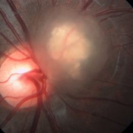
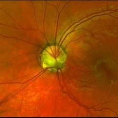
Fundus photograph of an 18-year-old woman with Leukemic optic neuropathy.
Photographer: Pedro Fernandes, Universidade Federal da Bahia
Condition/keywords: Leukemic optic neuropathy
-
 Optic Nerve Melanocytoma
Apr 3 2023 by Gustavo Aguirre Suarez
Fundus photograph of a 36-year-old female with a lesion dependent on the optic nerve head with subretinal extension, elevated, about 1.5 disc diameters, dark brown to black in color, involving more than three quarters of the neuroretinal ring towards the inferonasal area.
Photographer: Dr. Gustavo Aguirre-Suarez
Imaging device: Zeiss Visucam 500
Condition/keywords: melanocytic lesion, Melanocytoma
-
 Acute Syphilitic Posterior Placoid Chorioretinitis
Oct 20 2024 by César Adrián Gómez Valdivia, MD
Fundus autofluorescence image of an acute syphilitic posterior placoid chorioretinitis found in a HIV positive 28 YO male patient with suspected neurosyphilis. A beautiful butterfly autofluorescence pattern can be appreciated.
Photographer: @eyemissu2
Imaging device: California ICG OPTOS
Condition/keywords: acute syphilitic posterior placoid chorioretinitis
-
---thumb.jpg/image-square;max$300,300.ImageHandler) AMN
AMN
Aug 8 2013 by From the Collections of Thomas M. Aaberg, MD and Thomas M. Aaberg Jr., MD
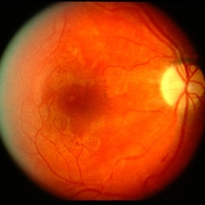
Wedge shaped brown-red lesions in the macula, left eye #2.
Condition/keywords: acute macular neuroretinopathy
-
 AMN (Acute Macular Neuroretinitis) #2
Apr 28 2019 by Niloofar Piri, MD
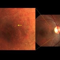
HD-OCT image of 53-year-old man who presented with a superior small paracentral scotoma for 1 month. He had very small hypopigmented area inferior to the fovea and hyporeflectivity on NIR image ( #1). OCT demonstrated a vertical hyper-reflective band extending from OPL to RPE. This form is type 2 AMN which is due to occlusion of deep capillary plexus.
Photographer: Niloofar Piri,MD
Condition/keywords: acute macular neuroretinitis, acute macular neuroretinopathy
-
 AMN (Acute Macular Neurortinitis)
Apr 28 2019 by Niloofar Piri, MD
HD-OCT image of 53-year-old man who presented with a superior small paracentral scotoma for 1 month. He had very small hypopigmented area inferior to the fovea and hyporeflectivity on NIR image ( #1). OCT demonstrated a vertical hyper-reflective band extending from OPL to RPE. This form is type 2 AMN which is due to occlusion of deep capillary plexus. #2
Condition/keywords: acute macular neuroretinitis, acute macular neuroretinopathy
-
 Astrocytic Hamartoma
Oct 10 2012 by Anat Loewenstein, MD
Five year-old girl came for regular eye examination with no complaints. Medical and ophthalmic history was unremarkable. On examination, visual acuity was 20/25 in both eyes. A Retinal Astrocytic Hamartoma was seen adjacent to the right optic nerve (picture). The rest of the eye examination was normal and no Lisch nodules were seen. The patient was referred for a pediatric neurologist examination and MRI scan of the brain.
Photographer: Galit Yair-Pur
Imaging device: ZEISS FF450 PLUS IR
-
 Central Serous Chorioretinopathy in Pregnancy (OS)
Apr 28 2024 by Vishal Agrawal, MD, FRCS,FACS,FASRS
30-year female with sudden loss of vision came for examination. She was in her first trimester of pregnancy. Examination revealed bilateral bullous NSD with subretinal fibrin s/o CSR.
Photographer: Dr Ayushi
Imaging device: Clarus 700
Condition/keywords: Central Serous Chorioretinopathy (CSR), neurosensory detachment of retina, pregnancy
-
 Choroidal Rupture
Apr 7 2021 by Priya Rasipuram Chandrasekaran, MBBS, DO, DNB, FRCS
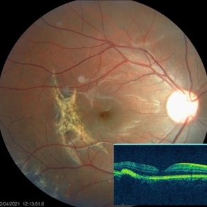
The fundus photo of a 24-year-old male shows crescent shaped choroidal rupture away from fovea and concentric to the optic disc following cricket ball injury. The corresponding optical coherence tomography shows disruption of the choriocapillaris, retinal pigment epithelium and Bruch’s membrane while the neurosensory retina remains intact. The fovea is not involved.
Condition/keywords: choroidal rupture
-
---thumb.jpg/image-square;max$300,300.ImageHandler) Diffuse Unilateral Subacute Neuroretinitis
Diffuse Unilateral Subacute Neuroretinitis
Feb 26 2013 by Henry J. Kaplan, MD
DUSN typical track and larva, subretinal.
Condition/keywords: diffuse unilateral subacute neuroretinitis (DUSN)
-
 DUSN (Diffuse Unilateral Subacute Neuroretinitis)
Sep 2 2016 by JEFFERSON R SOUSA, Tecg.º (Biomedical Systems Technology)
Patient female, 15-year-old, he entered the clinic with complaint of low vision, visual acuity without correction was 20/60 in the right eye and 20/30 in the left eye. In the ocular exam of retinografia, there was change in the epithelium macular pigment and a small larva juxtafoveal above.
Photographer: JEFFERSON R SOUSA - Study Center and Ophthalmological Research Dr. Andre M V Gomes, Institute Dr. Suel Abujamra São Paulo-Brazil
Imaging device: Topcon TRC-50 Dx - Angulation of field photo of 35 Degrees, flash 36, Digital system Imaginet
Condition/keywords: diffuse unilateral subacute neuroretinitis (DUSN), larva, uveitis
-
 Hypertensive optic neuropathy and choroidopathy right eye
Jan 11 2013 by Alex P. Hunyor, MD
Previous hypertensive optic neuropathy and choroidopathy, right eye. A young female who had a history severe pre-eclampsia. Note optic atrophy and multiple Elschnig spots.
Condition/keywords: hypertensive choroidopathy, hypertensive optic neuropathy
-
 Idiopathic retinal vasculitis, aneurysms and neuroretinitis
Apr 24 2022 by Aniruddha K Agarwal, MD
Ultra-wide field fundus fluorescein angiography (FFA) of the left eye from an asymptomatic, healthy 33-year-old woman who was referred to the retina clinic from a refractive surgery unit due to the presence of vascular anomalies and hard exudates in both eyes. FFA revealed the characteristic sacular aneurysms at the bifurcation of retinal arterioles in the posterior pole, together with microvascular anomalies and capillary closure peripherally.
Photographer: Julio J GONZALEZ-LOPEZ, MD, PhD, FEBO and Teresa GONZALEZ-LOMAS, RN
Imaging device: Optos California
Condition/keywords: IRVAN Syndrome, IUSG, neuroretinitis, retinal vasculitis, uveitis
-
 Idiopathic Retinal Vasculitis, Aneurysms, and Neuroretinitis (IRVAN)
Oct 16 2012 by S. Natarajan, MD, FASRS, FRCS (GLASGOW) , FICO, D.Sc, FELA
Fundus photograph of a young male with IRVAN Syndrome
Photographer: Prof. Dr. S. Natarajan
Condition/keywords: aneurysm, neuroretinitis, retinal vasculitis
-
 Idiopathic Retinal Vasculitis, Aneurysms, and Neuroretinitis (IRVAN)
Oct 16 2012 by S. Natarajan, MD, FASRS, FRCS (GLASGOW) , FICO, D.Sc, FELA
FFA photograph of a 28-year-old male with IRVAN Syndrome.
Photographer: Prof. Dr. S. Natarajan
Condition/keywords: aneurysm, neuroretinitis, retinal vasculitis
-
 Neuroretinitis
Jan 11 2013 by Alex P. Hunyor, MD
Leber's idiopathic stellate neuroretinitis, right eye.
Condition/keywords: neuroretinitis
-
 Optic Disc Edema With Macular Star
Jun 22 2013 by James A Eadie, MD
Fundus photograph montage of a 14-year-old girl with optic disc edema with macular star. Her laboratory work-up was negative for known causes. She improved from 20/200 to 20/40 with observation/an empirical course of doxycycline.
Photographer: Wendy Malmberg-Lorentz
Condition/keywords: neuroretinitis, optic disc edema
-
 Optic Nerve Pit Left Eye
Feb 15 2021 by Kim Barrett
A 14-year-old male presented with vision loss and VF defect. Patient was treated for presumed amblyopia with patching since age 4. He has had neurologic care for post traumatic skull fracture and brain bleed in 2012. Patient has a superior hemifield defect OS on HVF. IOP's WNL. There are vessels emanating from the optic pit OS. Patient is at risk of serous detachment. Current VA 20/20-2+2
Photographer: Kim Barrett C.O.A. Retina Specialist of Michigan, Grand Rapids, MI
Imaging device: Optos California
Condition/keywords: amblyopia, hemifield, Humphrey visual field, nerve, optic nerve pit, visual field defect
 Loading…