Search results (556 results)
-
 Toxoplasma Neuroretinitis (Jensen`s Disease)
Feb 25 2013 by Henry J. Kaplan, MD
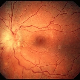
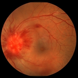
Toxoplasma neuroretinitis in the left eye of a patient with macular star formation, retinitis adjacent to the optic nerve head with disc swelling.
Condition/keywords: Jensen disease, ocular toxoplasmosis, toxoplasmosis
-
 Papillitis
May 2 2013 by Henry J. Kaplan, MD
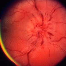
Anterior optic neuropathy or papillitis in the right eye; notice the blurred optic disc margin, engorged capillaries and flame shaped hemorrhages at the margin.
Condition/keywords: optic disc edema, optic disc swelling, papillitis
-
 AION With Ciliotretinal Artery Occlusion
May 2 2013 by Henry J. Kaplan, MD
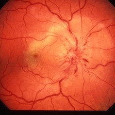
AION accompanied by partial CRAO which is visible as retinal edema and cherry red spot.
Condition/keywords: anterior ischemic optic neuropathy, central retinal artery occlusion (CRAO)
-
---thumb.jpg/image-square;max$300,300.ImageHandler) CMV Retinitis in a Patient with the Diagnosis of AIDS
CMV Retinitis in a Patient with the Diagnosis of AIDS
Feb 27 2013 by Henry J. Kaplan, MD
Color fundus photograph, right eye: CMV neuroretinitis (retinitis which begins from the arcades and is accompanied by hemorrhage and also has involved the optic nerve).
Condition/keywords: AIDS
-
---thumb.jpg/image-square;max$300,300.ImageHandler) Anterior Ischemic Optic Neuropathy
Anterior Ischemic Optic Neuropathy
Mar 29 2013 by Henry J. Kaplan, MD
Anterior Ischemic Optic Neuropathy; notice the typical pale optic disc swelling and faint splinter hemorrhages.
Condition/keywords: anterior ischemic optic neuropathy
-
 Idiopathic Retinal Vasculitis, Aneurysms, and Neuroretinitis (IRVAN)
Oct 16 2012 by S. Natarajan, MD, FASRS, FRCS (GLASGOW) , FICO, D.Sc, FELA
Fundus photograph of a young male with IRVAN Syndrome
Photographer: Prof. Dr. S. Natarajan
Condition/keywords: aneurysm, neuroretinitis, retinal vasculitis
-
---thumb.JPG/image-square;max$300,300.ImageHandler) Traumatic Optic Neuropathy
Traumatic Optic Neuropathy
Dec 9 2012 by Mallika Goyal, MD
Right eye of a 23-year-old gentleman 6 months following a road accident. Optic disc pallor with peripapillary chorioretinal scarring suggests traumatic optic neuropathy as the cause of optic atrophy.
Photographer: Mallika Goyal, MD, Apollo Health City, Hyderabad, India
Condition/keywords: traumatic optic neuropathy
-
 Wide-Field-OCT-montage
Jan 8 2018 by Netan Choudhry, MD, FRCS(C) FASRS
This is an SD-OCT montage image of a 55 year old male with optic neuropathy representing a wide-field OCT spanning 130 degrees.
Photographer: John Golding, Vitreous Retina Macula Specialists of Toronto
Imaging device: Heidelberg Spectralis OCT system
Condition/keywords: wide angle imaging
-
---thumb.JPG/image-square;max$300,300.ImageHandler) SLE retinopathy
SLE retinopathy
Nov 18 2013 by Mallika Goyal, MD
Occlusive retinitis in a lady with SLE; optic nerve head pallor suggestive of prior optic neuritis or ischaemic optic neuropathy.
Photographer: Mallika Goyal, MD, Apollo Health City, Hyderabad
Condition/keywords: systemic lupus erythematosus (SLE) retinopathy
-
---thumb.JPG/image-square;max$300,300.ImageHandler) Traumatic Optic Neuropathy
Traumatic Optic Neuropathy
Nov 28 2012 by Mallika Goyal, MD
Left eye of a 19-year-old boy 4 weeks following a road accident involving head injury as chest compression with lung contusion has visual acuity CF CF. There is disc pallor with surrounding retinal edema and hemorrhages suggestive of traumatic optic neuropathy.
Photographer: Mallika Goyal, MD, Apollo Health City, Hyderabad, India
Condition/keywords: traumatic optic neuropathy
-
 Sarcoid Optic Neuropathy with Retinal Hemorrhages
Oct 15 2012 by Jeffrey G. Gross, MD, FASRS
Sarcoid optic neuropathy with retinal hemorrhages, 20/50.
Condition/keywords: 20/50, autoimmunity, optic neuropathy, retinal hemorrhage, sarcoidosis
-
 AION
AION
Dec 19 2012 by Eric A. Postel, MD
fundus photograph of an elderly gentleman with AION
Condition/keywords: anterior ischemic optic neuropathy
-
 Choroidal Granuloma Secondary to Tuberculosis
Mar 14 2013 by Eduardo Torres-Porras, MD
OCT scan through the granuloma shows attachment of the retinal pigment epithelial-choriocapillaris layer and the neurosensory retina over the granuloma (“contact” sign), inflammatory retinal infiltrate in the deeper retinal layers and subretinal fluid.
Photographer: Eduardo Torres Porras, Laser y ultrasonido ocular de Puebla
Imaging device: Cirrus
Condition/keywords: optical coherence tomography (OCT), tubercular choroidal granuloma
-
 Optic Disc Drusen
Sep 21 2012 by Suber S. Huang, MD, MBA, FASRS
Fundus photograph of a 50-year-old woman with optic disc drusen complicated by anterior ischemic optic neuropathy
Condition/keywords: optic disc drusen
-
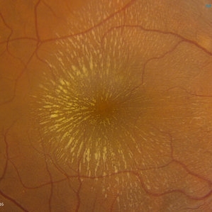
 Bilateral Macular Star
Mar 27 2014 by Jason S. Calhoun
Young female patient in with blurred vision in both eyes. VA is 20/40 in both eyes. Fundus photos show visible macular star centrally in both eyes. This is a result of Bilateral Neuroretinitis due to cat scratch.
Photographer: Jason S. Calhoun, Mayo Clinic Jacksonville, Department of Ophthalmology
Imaging device: TOPCON TRC 50-EX
Condition/keywords: macular star, neuroretinitis
-
 Idiopathic Retinal Vasculitis, Aneurysms, and Neuroretinitis (IRVAN)
Oct 16 2012 by S. Natarajan, MD, FASRS, FRCS (GLASGOW) , FICO, D.Sc, FELA
FFA photograph of a 28-year-old male with IRVAN Syndrome.
Photographer: Prof. Dr. S. Natarajan
Condition/keywords: aneurysm, neuroretinitis, retinal vasculitis
-
 Idiopathic Retinal Vasculitis, Aneurysms, and Neuroretinitis (IRVAN)
Oct 16 2012 by S. Natarajan, MD, FASRS, FRCS (GLASGOW) , FICO, D.Sc, FELA
Fundus photograph of a 28-year-old male with IRVAN Syndrome.
Photographer: Prof. Dr. S. Natarajan
Condition/keywords: aneurysm, neuroretinitis, retinal vasculitis
-
 Traumatic Optic Neuropathy
Nov 28 2012 by Mallika Goyal, MD
19-year-old status-post chest injury.
Condition/keywords: Purtscher's retinopathy, traumatic optic neuropathy, white centered retinal hemorrhage (Roth Spot)
-
---thumb.jpg/image-square;max$300,300.ImageHandler) Idiopathic Retinal Vasculitis, Aneurysms, and Neuroretinitis (IRVAN)
Idiopathic Retinal Vasculitis, Aneurysms, and Neuroretinitis (IRVAN)
Oct 16 2012 by S. Natarajan, MD, FASRS, FRCS (GLASGOW) , FICO, D.Sc, FELA
IRVAN
Condition/keywords: aneurysm, neuroretinitis, retinal vasculitis
-
 Idiopathic Retinal Vasculitis, Aneurysms, and Neuroretinitis (IRVAN)
Oct 16 2012 by S. Natarajan, MD, FASRS, FRCS (GLASGOW) , FICO, D.Sc, FELA
Fundus photograph of a 28-year-old male with IRVAN Syndrome.
Photographer: Prof. Dr. S. Natarajan
Condition/keywords: aneurysm, neuroretinitis, retinal vasculitis
-
 Choroidal Granuloma Secondary to Tuberculosis
Mar 14 2013 by Eduardo Torres-Porras, MD
OCT scan through the granuloma shows attachment of the retinal pigment epithelial-choriocapillaris layer and the neurosensory retina over the granuloma (“contact” sign), inflammatory retinal infiltrate in the deeper retinal layers and subretinal fluid.
Photographer: Eduardo Torres Porras
Imaging device: Cirrus
Condition/keywords: optical coherence tomography (OCT), tubercular choroidal granuloma
-
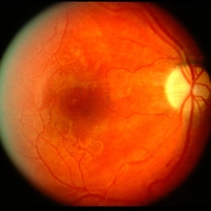
 Neurosensory Detachment with associated Pigment Epithelial Detachment
Oct 10 2012 by K. Bailey Freund, MD
Fundus photograph of a 39-year-old man with central serous chorioretinopathy noted to have a neurosensory detachment with associated pigment epithelial detachment.
Condition/keywords: central serous chorioretinopathy (CSCR), pigment epithelial detachment (PED)
-
 Cat Scratch
Feb 15 2017 by Hua Gao, MD, PhD, FASRS
A female patient of 57-year-old presented with neuroretinitis due to cat-scratch disease with positive Bartonella henselae antibodies. Two weeks after symptom onset she developed exudates in a "macular star" pattern.
Condition/keywords: cat scratch retinitis
-
 Hypertensive optic neuropathy and choroidopathy right eye
Jan 11 2013 by Alex P. Hunyor, MD
Previous hypertensive optic neuropathy and choroidopathy, right eye. A young female who had a history severe pre-eclampsia. Note optic atrophy and multiple Elschnig spots.
Condition/keywords: hypertensive choroidopathy, hypertensive optic neuropathy
-
 Idiopathic Retinal Vasculitis, Aneurysms, and Neuroretinitis (IRVAN)
Oct 16 2012 by S. Natarajan, MD, FASRS, FRCS (GLASGOW) , FICO, D.Sc, FELA
FFA photograph of a 28-year-old male with IRVAN Syndrome showing aneurysms around disc.
Photographer: Prof. Dr. S. Natarajan
Condition/keywords: aneurysm, neuroretinitis, retinal vasculitis
 Loading…