Search results (150 results)
-
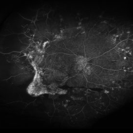
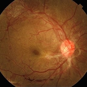 Extensive Neovascularization
Jul 21 2024 by César Adrián Gómez Valdivia, MD
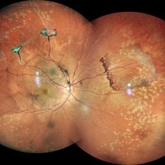
Macular Traction and Extensive Neovascularization found in a 66 year-old patient with history of uncontrolled Type 2 Diabetes Mellitus.
Photographer: Erika Paulina Ornelas Cazares
Imaging device: TOPCON TRC-50DX
Condition/keywords: diabetic retinopathy, neovascularization (NV)
-
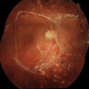 Preretinal Fibrosis
Jan 12 2024 by Virginia Gebhart
53 year old diabetic male with significant persistent ERM due to fibrotic NV superiorly. Possibly developing a tractional MH. Vitreous Hemorrhage secondary to traction on the fibrosis
Photographer: Virginia Gebhart
Imaging device: Topcon 50DX
Condition/keywords: epiretinal membrane, ERM, fibrosis, macular pseudohole, neovascularization (NV)
-
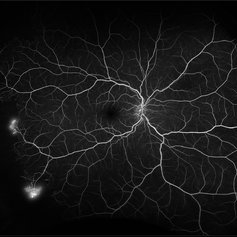
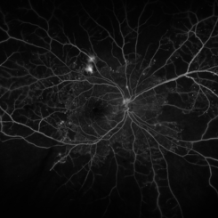
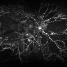
 Neovascularization in Posterior Uveitis
Jul 27 2023 by Zach Seim
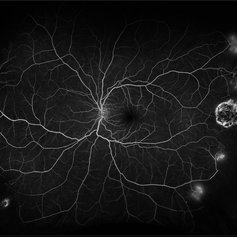
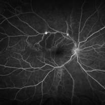
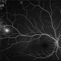
An ultra-widefield fluorescein angiogram of a 72 year old male with Posterior Uveitis and Neovascularization affecting the right eye. Patient's vision at the time of the image was Dcc 20/25. Dr. Korot states that the fluorescein angiogram shows patchy leakage throughout both eyes, with peripheral nonperfusion and secondary neovascularization. The patient was asked to get an extensive serological workup in an effort to identify any systemic autoimmune or infectious etiology as the cause for their severe inflammation.
Photographer: Zach Seim
Imaging device: OPTOS California
Condition/keywords: fluorescein angiogram (FA), FLUORESCEIN ANGIOGRAPHY, fluorescein leakage, neovascularization (NV), Optos, OPTOS CALIFORNIA, posterior uveitis, right eye, ultra-wide field imaging, ultra-widefield image
-
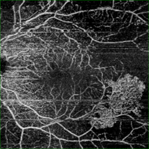
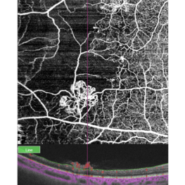 Retinal neovascularization
Feb 28 2023 by Nassim Alejandro Abreu Arbaje, MD
OCT and OCTa of a diabetic patient with severe PDR, showing the anatomical location and blood flow of neovessels
Photographer: Nassim Abreu, Centro de Oftalmologia y Glaucoma
Imaging device: Topcon Triton Plus
Condition/keywords: neovascularization (NV), OCT, OCT Angiography, PDR
-
 Proliferative Sickle Cell Retinopathy
Feb 1 2023 by Olivia Rainey
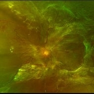
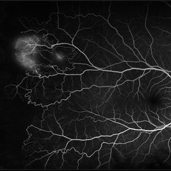
Ultra-widefield fluorescein angiography of a 25-year old male with Proliferative Sickle Cell Retinopathy affecting his right eye. Patient stated that he was born with Sickle disease (SC), and has yearly eye exams. He noted no vision concerns over the last year but has typically experienced sickle attacks about 1-2 per year. The physician noted that the fluorescein obtained showed peripheral nonperfusion affecting the patient's nasal and temporal retina as well as neovascularization affecting his left eye more than his right. He recommended pan retinal photocoagulation in his left eye for his temporal and nasal retina, as as well as his right eye following.
Photographer: Olivia Rainey, OCT-C, COA
Imaging device: Optos California
Condition/keywords: early phase, fluorescein angiogram (FA), fluorescein leakage, neovascularization (NV), non-perfusion, proliferative retinopathy, right eye, sickle cell retinopathy, ultra-wide field imaging, ultra-widefield image
-
 Proliferative Sickle Cell Retinopathy
Feb 1 2023 by Olivia Rainey
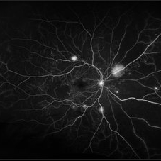
Ultra-widefield fluorescein angiography of a 25-year old male with Proliferative Sickle Cell Retinopathy affecting his left eye. Patient stated that he was born with Sickle disease (SC), and has yearly eye exams. He noted no vision concerns over the last year but has typically experienced sickle attacks about 1-2 per year. The physician noted that the fluorescein obtained showed peripheral nonperfusion affecting the patient's nasal and temporal retina as well as neovascularization affecting his left eye more than his right. He recommended pan retinal photocoagulation in his left eye for his temporal and nasal retina, as as well as his right eye following.
Photographer: Olivia Rainey, OCT-C, COA
Imaging device: Optos California
Condition/keywords: early phase, fluorescein angiogram (FA), fluorescein leakage, left eye, neovascularization (NV), proliferative retinopathy, sickle cell retinopathy, ultra-wide field imaging, ultra-widefield image
-
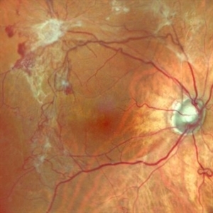
 Diabetic Traction Detachment of Retina
Sep 28 2022 by Chloe Hanifan
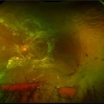
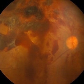
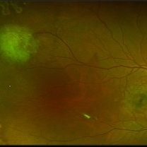
Ultra-widefield pseudo color fundus photograph of a 50-year-old female with a Diabetic Traction Detachment of Retina affecting her left eye. The patient was unable to proceed with surgery due to other health issues in May of 2022, when she presented in the office in September of 2022, a guarded prognosis given chronicity and associated ischemia. The patient was LP at the time of the September appointment.
Photographer: Chloe Hanifan
Imaging device: Optos California
Condition/keywords: Diabetes, diabetic traction detachment, fundus photography, left eye, neovascularization (NV), Optos, proliferative diabetic retinopathy (PDR), pseudocolor, ULTRA WIDE FIELD
-
 Diabetic Traction Detachment of Retina
Sep 28 2022 by Chloe Hanifan
Ultra-widefield pseudo color fundus photograph of a 50-year-old female with a Diabetic Traction Detachment of Retina affecting her left eye. The patient was unable to proceed with surgery due to other health issues in May of 2022, when she presented in the office in September of 2022, a guarded prognosis given chronicity and associated ischemia. The patient was LP at the time of the September appointment.
Photographer: Chloe Hanifan
Imaging device: Optos California
Condition/keywords: diabetes, diabetic traction detachment, fundus photography, left eye, neovascularization (NV), Optos, proliferative diabetic retinopathy (PDR), pseudocolor, ULTRA WIDE FIELD
-
 Neovascular vessels
Sep 22 2022 by Filip Kecer
Multicolor widefield scan of a 16-year-old girl with a neovascularization from disc to vitreous space
Photographer: Filip Kecer, National Institute of Childrens Diseases
Imaging device: Spectralis, Heidelberg Engineering
Condition/keywords: neovascularization (NV), neovascularization at the disc, uveitis, vitreous
-
 Neovascularization in a combined CRAO/BRVO
Feb 21 2022 by Maxwell J Wingelaar, MD
60-year-old male with neovascularization from a combined CRAO/BRVO
Condition/keywords: neovascularization (NV)
-
 Monocular Proliferative Diabetic Retinopathy
Sep 8 2021 by VERONICA ADRIANA ROMERO- MORALES, MD
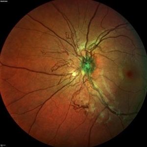
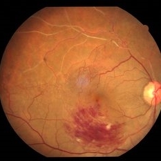
Fundus photograph of a 37-year-old woman with proliferative diabetic retinopathy and subhyaloid hemorrhage, 1 week of evolution.
Photographer: Belgica Copado Andrade
Imaging device: Cobra HD
Condition/keywords: neovascularization (NV), proliferative diabetic retinopathy (PDR), subhyaloid hemorrhage, thickening of the posterior hyaloid, vitreous blood
-
 Neovascularization in a Case of Idiopathic Vasculitis
Aug 18 2021 by ASRS Staff
Right eye, Wide field photograph Motage of 37 year-old male, having idiopathic vasculitis status post PRP. Patient's systemic evaluation was done, and everything was within normal limits.
Imaging device: Nidek Mirante
Condition/keywords: Idiopathic vasculitis, montage, neovascularization (NV), pan-retinal photocoagulation (PRP)
-
 Battle of BRVOs: Old vs New
Jul 30 2021 by Gayathri Mohan
Color fundus photograph of a 48-year-old showing an inferno temporal BRVO. Old BRVO with neovascularization seen super-temporally.
Photographer: Dr. Gayathri Mohan
Imaging device: Canon
Condition/keywords: branch retinal vein occlusion (BRVO), cystoid macular edema (CME), neovascularization (NV)
-
 Neovascular Glaucoma Tree
Jun 23 2021 by Ana Karen Lopez, MD
Anterior segment photography of an 54-year-old man with neovascular glaucoma.
Photographer: Ana Karen López-Vázquez, MD
Condition/keywords: angle neovascularization, anterior chamber, cataract with neovascularization, neovascular glaucoma, neovascularization (NV), neovascularization of iris (NVI)
-
 Sickle Cell Retinopathy
Feb 15 2021 by Kim Barrett
24-year-old female with Sickle Cell Retinopathy, stage 3. She confirms she has the trait as well as her grandmother, mother and a sibling. She has seafan neovascularization superotemporal OD. Current VA is 20/20. Photo is pre-PRP laser with areas of non-profusion temporally.
Photographer: Kim Barrett C.O.A. Retina Specialist of Michigan, Grand Rapids, MI
Imaging device: Optos California
Condition/keywords: neovascularization (NV), pan-retinal photocoagulation (PRP), sickle cell retinopathy, stage 3, trait
-
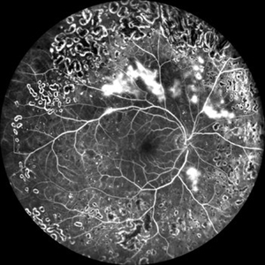
 Proliferative Diabetic Retinopathy
Jan 29 2021 by Olivia Rainey
Ultra-widefield fluorescein angiogram of a 65-year-old male with proliferative diabetic retinopathy affecting his right eye. The patient's diabetic retinopathy has progressed significantly since he was last seen in 2014. It was recommended to begin antiVEGF to control DME followed by laser treatment OU.
Photographer: Olivia Rainey, OCT-C, COA
Imaging device: Optos California
Condition/keywords: anti-VEGF, diabetes, diabetic macular edema, fluorescein angiogram (FA), fluorescein leakage, neovascularization (NV), neovascularization elsewhere (NVE), non-perfusion, Optos, proliferative diabetic retinopathy (PDR), ultra-wide field imaging
-
 Proliferative Sickle Cell Retinopathy
Jan 29 2021 by Olivia Rainey
Ultra-widefield fluorescein angiogram of a 24-year-old female with proliferative sickle cell retinopathy affecting her right eye. The physician's interpretation of the fluorescein shows seafan neovascularization superotemporally, AV anastomeses, and good peripheral laser. He performed scatter PRP OD on 12/2/2020 to nonperfusion in temporal far periphery. The patient's 12/2020 Hb electrophoresis came back showing Hb SC (rather than sickle cell trait). Patient was born at full term, but she reports that her mother used drugs while pregnant with the patient. The patient also mentioned that her niece has full sickle cell disease and her grandmother, mother, and sibling all have condition on the sickle cell spectrum.
Photographer: Olivia Rainey, OCT-C, COA
Imaging device: Optos California
Condition/keywords: fluorescein angiogram (FA), fluorescein leakage, neovascularization (NV), neovascularization elsewhere (NVE), Optos, sea fan, sickle cell retinopathy
-
 Proliferative Sickle Cell Retinopathy
Jan 29 2021 by Olivia Rainey
Ultra-widefield fundus photograph of a 24-year-old female with proliferative sickle cell retinopathy affecting her right eye. He performed scatter PRP OD on 12/2/2020 to nonperfusion in temporal far periphery. The patient's 12/2020 Hb electrophoresis came back showing Hb SC (rather than sickle cell trait). Patient was born at full term, but she reports that her mother used drugs while pregnant with the patient. The patient also mentioned that her niece has full sickle cell disease and her grandmother, mother, and sibling all have condition on the sickle cell spectrum.
Photographer: Olivia Rainey, OCT-C, COA
Imaging device: Optos California
Condition/keywords: fundus photograph, laser photocoagulation, neovascularization (NV), neovascularization elsewhere (NVE), Optos, pseudocolor, sea fan, sickle cell retinopathy
-
 Severe Proliferative Diabetic Retinopathy with Asteroid Hyalosis
Aug 25 2020 by Olivia Rainey
Ultra-widefield fluorescein angiogram of a 47-year-old male with severe prolifterative diabetic retinopathy with very extensive neovascularization with fibrosis and traction affecting his right eye. The patient also has asteroid hyalosis affecting the eye. He was diagnosed with Type 1 diabetes in the late 1970s. The patient's vision sc20/100 PH20/70-2. He received treatment of panretinal photocoagulation following the angiogram.
Photographer: Olivia Rainey, OCT-C, COA
Imaging device: Optos California
Condition/keywords: asteroid hyalosis, FA early phase, fibrotic neovascularization, fluorescein angiogram (FA), hyperfluorescence, neovascularization (NV), ultra-wide field imaging
-
 PDR with Ischemia
Jul 7 2020 by Stephanie Burke
Early frame of a 45-year-old male with Type II diabetes.
Photographer: Stephanie Burke, CRA, OCT-C
Condition/keywords: FA early phase, ischemia, microaneurysms, neovascularization (NV), proliferative diabetic retinopathy (PDR), ultra-wide field imaging, venous beading
-
 Neovascularization in PDR
Jun 10 2020 by Manish Nagpal, MD, FRCS (UK), FASRS
Neovascular fronds in PDR
Photographer: Gayathri Mohan, Retina Foundation
Imaging device: NIDEK SLO MIRANTE
Condition/keywords: neovascularization (NV), proliferative diabetic retinopathy (PDR)
-
 Fluorescein Angiography Revealing Neovascularization in Lasered PDR
Jun 10 2020 by Manish Nagpal, MD, FRCS (UK), FASRS
Fluorescein angiography revealing neovascularization in lasered PDR.
Photographer: gayathri mohan
Imaging device: nidek slo mirante
Condition/keywords: fundus photograph, neovascularization (NV), proliferative diabetic retinopathy (PDR)
-
 OCT Angiography- PDR
May 11 2020 by Gayathri Mohan
OCT angiography image of the superficial plexus showing neovascularisation infero-temporal to macula. Avascular areas are seen temporally.
Photographer: Gayathri Mohan, Retina Foundation
Imaging device: Mirante, Nidek
Condition/keywords: neovascularization (NV), optical coherence tomography (OCT), proliferative diabetic retinopathy (PDR)
-
 Proliferative Diabetic Retinopathy
May 11 2020 by Gayathri Mohan
Color fundus photograph of a patient with PDR, showing neovascularisation infero-temporal to macula.
Photographer: Gayathri Mohan, Retina Foundation
Imaging device: Mirante, Nidek
Condition/keywords: neovascularization (NV), optical coherence tomography (OCT), proliferative diabetic retinopathy (PDR)
-
 Proliferative Diabetic Retinopathy
Apr 29 2020 by Stephanie Burke
Ultra-wide FA image of a 58-year-old man with DM.
Photographer: Stephanie Burke, CRA, OCT-C
Condition/keywords: ischemia, leakage, microaneurysms, neovascularization (NV), proliferative diabetic retinopathy (PDR), ultra-wide field imaging
 Loading…