Search results (3038 results)
-
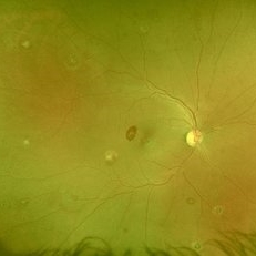
 CRAO With Cilio-retinal Sparing-MMI
Jun 25 2025 by Shivankar Sen
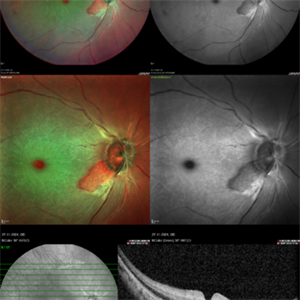
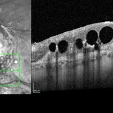
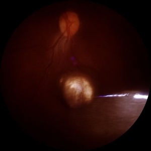
A 41 year old male came with complaints of Right eye blurring of vision since a day associated with watering and redness. He had no systemic illness, though gave a history of fall from bike 1 month back at the time of which he had blunt force trauma to the right side of the face. BCVA was 3/60, less than N36 in the right eye and 6/6, N6 in the left eye. Right eye had Marcus Gunn Pupil with clear lens, Left eye was within normal limits. IOP was normal; 16 in OD and 18 in OS. Retina evaluation revealed CRAO in the right eye with cilio-retinal artery sparing. Left eye was unremarkable Image Details Left to Right (Top 2 rows) Multicolor Reflectance Image (Blue-green enhanced 55 degree) revealing cilioretinal spared retinal stroma and a characteristic Cherry Red Spot; Green Reflectance showing corresopnding dark gray area with spared perfusion and black spot consistent with Cherry Red Spot on multicolor 2nd Row - 35 degree image (Multicolor Standard Reflectance and Green Reflectance) 3rd Row - SD-OCT revealing acute moderate CRAO findings with Middle retinal layer opacification and prominent middle limiting membrane (p-MLM) sign; Inner retinal layer opacification and prominent retinal pigment epithelium at the fovea with Diminished inner retinal layer stratification
Photographer: Gayathri M S
Imaging device: Heidelberg Spectralis HRA+OCT
Condition/keywords: CRAO with cilioretinal sparing, multicolor, multimodal imaging, OCT biomarkers, reflectance
-
 Berlins Edema - Multimodal Imaging
Jun 25 2025 by Shivankar Sen
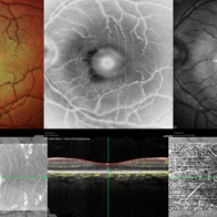
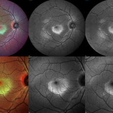
A 22 year old female came with history of injury to her left eye with a badminton racquet butt cap an hour before presentation On examination, she was found to have right eye 6/6;N6 vision and within normal limits, left eye 6/9;N6 vision, cells1+ in the anterior chamber, brisk pupillary response, no vitreous reaction and sub-clinical berlin's edema at the posterior pole. Multimodal imaging revealed frank boundaries of Berlin's edema more pronounced in the nasal parafoveal region. Figure details Top (Left to Right) Multicolor Reflectance showing bright yellow ring surrounding the perifovea; Blue Reflectance (Black on white contrast) showing corresponding black ring; Green Reflectance showing a characteristic white ring (all pronounced nasally); Bottom (Left-Right) Transverse structural OCT enface image showing white ring consistent with edema OCTA inner layer segmentation from ILM to GCL Transverse corresponding OCTA revealing faint hypo ring within perifoveal capillary bed
Photographer: Gayathri M S
Imaging device: Heidelberg Spectralis HRA+OCT
Condition/keywords: blue reflectance, En Face OCTA, enface imaging, multicolor, oct, reflectance
-
 Berlins
Jun 25 2025 by Shivankar Sen
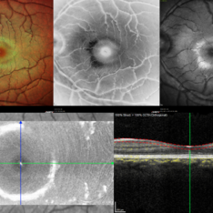
A 22 year old female came with history of injury to her left eye with a badminton racquet butt cap an hour before presentation On examination, she was found to have right eye 6/6;N6 vision and within normal limits, left eye 6/9;N6 vision, cells1+ in the anterior chamber, brisk pupillary response, no vitreous reaction and sub-clinical berlin's edema at the posterior pole. Multimodal imaging revealed frank boundaries of Berlin's edema more pronounced in the nasal parafoveal region. Figure details Top (Left to Right) Multicolor Reflectance showing bright yellow ring surrounding the perifovea; Blue Reflectance (Black on white contrast) showing corresponding black ring; Green Reflectance showing a characteristic white ring (all pronounced nasally); Bottom (Left-Right) Transverse structural OCT enface image showing white ring consistent with edema OCTA inner layer segmentation from ILM to GCL
Photographer: Gayathri M S
Imaging device: Heidelberg Spectralis HRA+OCT
Condition/keywords: blue reflectance, En Face OCTA, multicolor
-
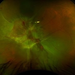
 Active Multi Focal Choroiditis
Jun 21 2025 by Moazzam Parvez
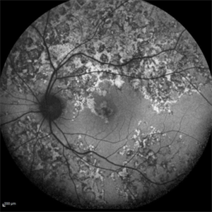
Auto fluorescence image of a 28 year old gentleman with active multifocal choroiditis in his left eye and healed choroiditic patches in the right eye.
Photographer: Moazzam Parvez , Netralayam , Kolkata
Imaging device: Heidelberg Spectralis
Condition/keywords: active, multifocal choroiditis
-
 Subretinal PFO
Jun 18 2025 by Korey Starkey
86-year-old patient had history for retinal detachment surgery x2 and intraocular injections for AMD performed elsewhere. Left eye has PVR developing and subretinal PFO. Due to guarded vision, opting to defer any further treatment at this time.
Photographer: Korey Starkey
Imaging device: Heidelberg
Condition/keywords: AMD, Heidelburg Spectralis, OCT, PFO, PVR, retinal detachment, silicone oil
-
 Berlin's Edema
Jun 12 2025 by Shivankar Sen
A 22 year old male came with history of sports injury to the right eye with the nose of shuttlecock while playing badminton. On examination, right eye anterior segment shows conjunctival congestion with brisk pupillary reaction and quiet anterior chamber. His best corrected visual acuity was 6/12; N6 in the right eye and 6/6; N6 in the left eye. Retinal examination revealed OD Berlin's Edema, OS within normal limits. Image Description (From Left to Right) Multicolor Reflectance (Blue-Green Enhanced) shows well defined yellowish discoloration Green reflectance and blue reflectance show corresponding whitish discoloration at the area of edema
Photographer: Dr. Shivankar Sen
Imaging device: Heidelberg Spectralis HRA+OCT
Condition/keywords: Shuttlecock Injury
-
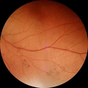
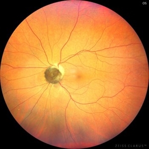
 Commotio Retinae
Jun 10 2025 by CUI YUELING
The patient presented 2 hours after sustaining a left eye injury caused by a stick. Visual acuity in the left eye was 0.2 without improvement upon correction, and intraocular pressure measured 15 mmHg. Examination of the anterior segment revealed ciliary conjunctival injection accompanied by patchy subconjunctival hemorrhage. The corneal surface remained smooth, and the anterior chamber was deep with hyphema characterized by blood-tinged aqueous humor predominantly settled inferiorly. The pupil was slightly irregular, approximately 3 mm in diameter, with a superotemporal notch; pupillary light reflex was intact. The lens appeared clear. Fundus examination showed well-defined optic disc margins with normal coloration and a cup-to-disc ratio of 0.2. Retinal arteries and veins were normally distributed with an artery-to-vein ratio of 2:3. At the posterior pole, the foveal reflex exhibited concentric ripple-like changes centered on the fovea, accompanied by localized pigment attenuation and reduced reflex intensity. Irregular reflectivity was noted in the superotemporal and inferotemporal nerve fiber layers.
Photographer: Yueling Cui
Imaging device: Zeiss Clarus 500
Condition/keywords: commotio retinae
-
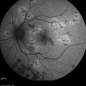
 Extensive Macular Atrophy with Pseudodrusen EMAP OS
Jun 5 2025 by Rogerio N Shinsato, MD, PhD
Extensive Macular Atrophy with Pseudodrusen Patient with EMAP associated with rheumatic fever and benzathine penicillin use. Left eye
Photographer: Rogério N Shinsato
Imaging device: Canon CX-1
Condition/keywords: EMAP
-
 Tuberculoma
Jun 4 2025 by Paulina Araujo
The 55-degree central fundus photograph of the left eye reveals an elevated, nodular whitish choroidal lesion along the inferior temporal arcade, consistent with a tuberculoma.
Photographer: Paulina D.Araujo Martínez, Asociación para Evitar la Ceguera en México I.A.P., Hospital Dr Luis Sánchez Bulnes.
Condition/keywords: Choroidal-tuberculoma
-
 Myelinated Nerve Fibers
Jun 4 2025 by Paulina Araujo
The 55-degree central fundus photograph of the left eye reveals myelination of the nerve fiber layer along the inferior nasal arcade.
Photographer: Paulina D.Araujo Martínez, Asociación para Evitar la Ceguera en México I.A.P., Hospital Dr Luis Sánchez Bulnes.
Condition/keywords: myelinated nerve fibers
-
 Bear Tracks (CHRPE)
Jun 4 2025 by Paulina Araujo
The 55-degree fundus photograph of the left eye shows bear tracks along the inferior temporal arcade.
Photographer: Paulina D.Araujo Martínez, Asociación para Evitar la Ceguera en México I.A.P., Hospital Dr Luis Sánchez Bulnes.
Condition/keywords: bear tracks, congenital hypertrophy of the retinal pigment epithelium (CHRPE)
-
 Choroidal Rupture
Jun 4 2025 by Paulina Araujo
The 55-degree central fundus photograph of the left eye reveals a choroidal rupture in the nasal parafoveal area secondary to blunt ocular trauma.
Photographer: Paulina D.Araujo Martínez, Asociación para Evitar la Ceguera en México I.A.P., Hospital Dr Luis Sánchez Bulnes.
Condition/keywords: choroidal rupture
-
 Tessellated Fundus
Jun 4 2025 by Paulina Araujo
The 55-degree central fundus photograph of the left eye reveals a tessellated fundus appearance consistent with high myopia.
Photographer: Paulina D.Araujo Martínez, Asociación para Evitar la Ceguera en México I.A.P., Hospital Dr Luis Sánchez Bulnes.
Condition/keywords: Tessellated fundus
-
 Tractional/Rhegmatogenous Retinal Detachment
May 29 2025 by Jenn Geelan
48 year-old male with a combined Tractional/ Rhegmatogenous retinal detachment with NVD and Persistent Fetal Vasculature in the left eye.
Photographer: Jenn Geelan, Retina-Vitreous Surgeons of CNY
Condition/keywords: fundus photograph, Neovascularisation at the Disc (NVD), OPTOS CALIFORNIA, persistent fetal vasculature (PFV), retinal detachment, rhegmatogenous retinal detachment, tractional retinal detachment
-
 Active multifocal choroiditis
May 26 2025 by Moazzam Parvez
Auto fluorescence photograph of an 43 year old man with active choroiditic lesion present in the left eye with recurrence
Photographer: Dr Moazzam Parvez , Netralayam , Kolkata
Imaging device: Heidelberg Spectralis
Condition/keywords: active choroididtis, choroiditi
-
 Leukemic Infiltrate
May 11 2025 by Hemanth Murthy, MBBS, MD, FASRS
43 year male patient presented with blurring of vision in right eye since 3 days. Vision 6/12 and left eye vision was 6/6. Haematological workup showed Hemoglobin -10g/dl, WBC count 276440 cells/cu.mm Smear showed large immature myeloid cells.
Photographer: Mr Veda Vyas
Condition/keywords: Acute myeloid leukaemia with Roth spots and leukaemia infiltrates
-
 White and Red Spots- Roth Spots and Leukemic Infiltrates in Acute Myeloid Leukemia
May 11 2025 by Hemanth Murthy, MBBS, MD, FASRS
43 year male patient presented with blurring of vision in right eye since 3 days. Vision 6/12 and left eye vision was 6/6. Haematological workup showed Hemoglobin -10g/dl, WBC count 276440 cells/cu.mm Smear showed large immature myeloid cells.
Photographer: Mr Veda Vyas
Condition/keywords: Acute myeloid leukaemia with Roth spots and leukaemia infiltrates
-
 Repaired Retinal Detachment
May 7 2025 by Kimberly Wakester
Optomap RGB montage of an 56-year-old woman with a repaired retinal detachment with scleral buckle and cryotherapy in the left eye. Patient remains stable s/p Vitreo-retinal surgery in 2007. Patient is to return in 1 year for follow up exam with repeat imaging.
Photographer: Kimberly Wakester, COA, OCT-C
Imaging device: Optos California
Condition/keywords: cryotherapy, repaired RD, scleral buckle
-
 Optic Nerve Melanocytoma
May 4 2025 by KANWALJEET HARJOT MADAN, M.S. (Ophthalmology); FAICO (Vitreous - Retina)
This is a fundus picture of a young 42-year male who visited for a routine eye exam. His BCVA was 20/20 in both eyes. Anterior segment examination was normal. His left eye showed grey-black pigmentation at the infero-nasal margin of the optic disc. Fundus of the right eye was normal. The patient was diagnosed to have optic disc melanocytoma on multimodal imaging and was advised regular follow-up. Optic nerve melanocytoma is typically a benign tumor made up of melanocytes and melanin. It can grow, but rarely transforms into a malignancy. Patients with Optic Nerve Melanocytoma should be periodically examined for evidence of growth, loss of visual field and optic nerve compression.
Photographer: Dr. Kanwaljeet Harjot Madan, Thind Eye Hospital, Jalandhar City (Punjab) INDIA.
Imaging device: Zeiss Fundus Camera
Condition/keywords: melanocytoma, melanoma, optic nerve
-
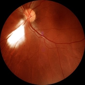
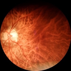
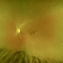
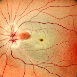 CRAO Sparing Cilioretinal Artery
May 1 2025 by Tejaswita Verma
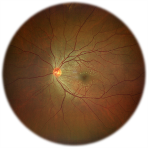
Fundus photo of a middle aged male with 6/60 vision in left eye showing CRAO partially sparing cilioretinal artery.
Photographer: Dr. Tejaswita Verma
Imaging device: MIRANTE
Condition/keywords: cilioretinal sparing, CRAO
-
 Retinal Macroaneurysm (Left Eye)
Apr 29 2025 by Daniela Bogenschutz
72 year-old female has visual complaints of central vision changes ongoing for 4 days. Patient was acutely symptomatic with an intraretinal hemorrhage due to the retinal macroaneurysm. We had a fun little laugh as this retinal macroaneurysm form a shape of a tick in her left eye. This photo is a side-by-side of the color photos and the autofluorescence done. She is being treated by her general doctor for elevated blood pressure.
Photographer: Daniela Bogenschutz, OSC; Retina Consultants of Carolina, P.A.
Imaging device: Optos
Condition/keywords: retinal macroaneurysm
-
 Retinitis Pigmentosa with Macular Hole with Posterior Subcapsular Cataract
Apr 28 2025 by Malvika Singh
Fundus photograph of the left eye of a 31 year old with retinitis pigmentosa, showing the shadow of posterior subcapsular cataract over the fundus.
Photographer: Dr Malvika Singh, Retina Foundation, Ahmedabad, India
Imaging device: Mirante SLO/OCT
Condition/keywords: posterior subcapsular cataract, retinitis pigmentosa
-
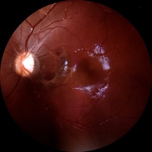
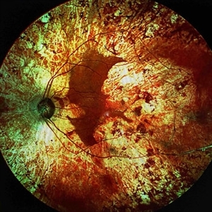
 Extensive Chorioretinal Scarring With Partial Macular Sparing
Apr 22 2025 by Maxwell J Wingelaar, MD
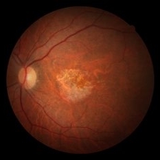
Fundus autofluorescence of extensive chorioretinal scarring in the left eye.
Photographer: Killian Roberts
Imaging device: Heidelberg Spectralis AF
Condition/keywords: chorioretinal atrophy, chorioretinal inflammations
-
 Extensive Chorioretinal Scarring with Partial Macular Sparring
Apr 22 2025 by Maxwell J Wingelaar, MD
A multicolor photo showing chorioretinal scarring with partial macular sparing in the left eye.
Photographer: Killian Roberts
Imaging device: Heidelberg Spectralis Multicolor Photo
Condition/keywords: chorioretinal atrophy, chorioretinal inflammations
 Loading…