Search results (3502 results)
-
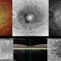
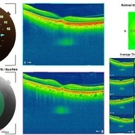 Adult Foveomacular Dystrophy
Aug 21 2025 by Aditya S Kelkar, MS, FRCS, FASRS,FRCOphth
Left eye OCT of a 56 year old female with complaints of gradual painless blurring of vision, aggravating on near work.
Photographer: Dr. Muskan Mangal
Condition/keywords: adult foveomacular dystrophy, OCT
-
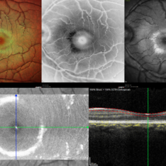
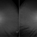 Dystrophy of the Retinal Pigment Epithelium
Aug 21 2025 by Aditya S Kelkar, MS, FRCS, FASRS,FRCOphth
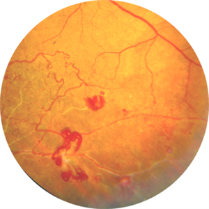
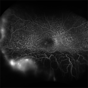
Both eyes autofluorescence imaging on Optos of a 56 year old female with complaints of gradual painless blurring of vision, aggravating on near work. Her BCVA for distance vision is 6/12 and 6/9 on snellens charting for Right and Left eye respectively. What could be the exact pathology or diagnosis? Kindly discuss and suggest.
Photographer: Dr. Muskan Mangal
Condition/keywords: autofluorescence imaging, Dystrophy of the Retinal Pigment Epithelium, macula lesion
-
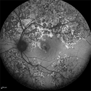
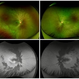 Goldmann-Favre Syndrome
Aug 19 2025 by Debarun Sharma
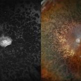
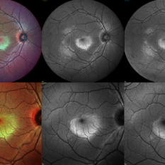
Fundus photograph of a 17 year-old female showing circumferential nummular opacities surrounding the vascular arcades. Fundus autoflourescence shows hypo-autoflourescent circumferential opacities with hyper-autoflourescent ring surrounding macula. Left eye also shows hyper-autoflourescent lesion on the optic nerve head suggestive of astrocytic hamartoma. ERG showed reduced cone response with extinguished rod response. OCT showed schisis of macular area. These features are suggestive of Goldmann-Favre Syndrome.
Photographer: Dr. Debarun Sharma, Sri Sankardeva Nethralaya, Guwahati
Imaging device: Optos
Condition/keywords: Goldmann-Favre Syndrome
-
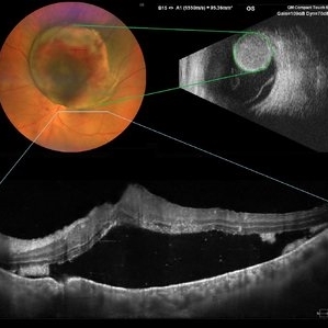 Choroidal Melanoma
Aug 19 2025 by JEFFERSON R SOUSA, Tecg.º (Biomedical Systems Technology)
A 54-year-old woman with progressive visual acuity loss in her left eye was admitted to the institution with a significant elevated lesion in the upper arch with macular involvement, confirmed by wide-angle fundus photography, ultrasound, and optical coherence tomography.
Photographer: JEFFERSON ROCHA DE SOUSA - Retinal Department at Lens Oftalmologia, Sao Paulo-Brazil
Imaging device: Clarus 700 - Zeiss, composite of four 135 degree images.
Condition/keywords: melanoma
-
 Retinal Detachment
Aug 12 2025 by Kimberly Wakester
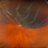
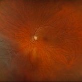
Optomap RGB of a 63-year-old man with a bullous overhanging Retinal Detachment with superonasal tuft-associated tear, macula detached in the left eye. Surgery was recommended. Patient is to continue follow up care post operatively.
Photographer: Kimberly Wakester, COA, OCT-C, Retina Consultants of Carolina
Imaging device: Optos California
Condition/keywords: bullous retinal detachment, left eye, Mac off
-
 Retinal Detachment with Multiple Breaks
Aug 12 2025 by Kimberly Wakester
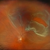
Optomap RGB of a 59-year-old man with a retinal detachment with multiple breaks in the left eye. Surgery was recommended. Patient is to continue follow up care post operatively.
Photographer: Kimberly Wakester, COA, OCT-C, Retina Consultants of Carolina
Imaging device: Optos California
Condition/keywords: lattice degeneration, left eye, Retinal Detachment with Multiple Breaks
-
 Macular Star
Aug 6 2025 by Tadeo Blanco
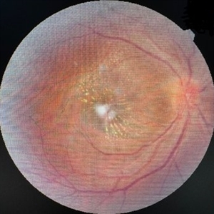
Fundus photograph of a 44 year-old woman with a macular star. She has type 2 diabetes, close living with 8 cats. Presents with decreased visual acuity in the right eye with 1 month of evolution. Refers a febrile episode one month prior. Visual acuity: right eye 20/200, left eye 20/25. Ishihara test: right eye 4/10, left eye 10/10. Bartonella henselae IgG: positive.
Photographer: R. Tadeo Blanco-Nunez
Condition/keywords: macular star, ocular bartonellosis
-
 Horseshoe Retinal Tear
Aug 6 2025 by Korey Starkey
80 year-old patient presented with HSRT without detachment in the left eye and macula-off detachment in the right eye. Scheduled patient for prompt surgical repair OD and same day laser retinopexy OS to reduce risk of retinal detachment.
Photographer: Korey Starkey
Imaging device: Optos
Condition/keywords: color fundus photograph, fundus photography, horseshoe tear, Optos
-
 Desert Rose
Aug 4 2025 by KANWALJEET HARJOT MADAN, M.S. (Ophthalmology); FAICO (Vitreous - Retina)
This is fundus picture of left eye of a 54 years male depicting Supero Temporal Branch Retinal Vein Occlusion with collaterals and neo vascularization elsewhere.
Photographer: Dr. Kanwaljeet Harjot Madan, Thind Eye Hospital, Jalandhar City (Punjab). INDIA.
Imaging device: Zeiss Fundus Camera
Condition/keywords: Neo Vascularization, Supero Temporal Branch Retinal Vein Occlusion
-
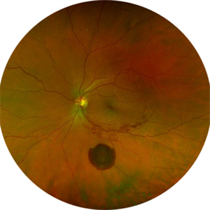
 Torpedo Maculopathy
Aug 2 2025 by Gabriel Costa Andrade, PhD
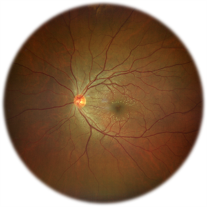
Color fundus image of a 41-year-old male ina routine examination with 20/20 vision in both eyes presenting with torpedo-shaped macula lesion in the temporal macular area of left eye.
Photographer: Gabriel Andrade
Condition/keywords: macula, Maculopathy
-
 Anastomosis
Jul 29 2025 by Drew Mitchell
3x3 OCT-Angiography Full Depth Color Coded of a left eye with Macular Telangiectasia Type 2
Photographer: Drew Mitchell, OCT-C
Imaging device: Zeiss Cirrus 5000
Condition/keywords: chorioretinal anastomosis, macular telangiectasia type 2, retinochoroidal anastomosis
-
 Retinal detachment with Single Break
Jul 18 2025 by Kimberly Wakester
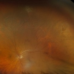
Optomap RGB of a 62-year-old man with a retinal detachment with a single break in the left eye. Patient has a previously treated HSRT in the left eye. Surgery was recommended. Patient is to continue follow up care post operatively.
Photographer: Kimberly Wakester, COA, OCT-C
Imaging device: Optos California
Condition/keywords: RD, retinal tear
-
 Pigmentary Retinal Dystrophy
Jul 18 2025 by Kimberly Wakester
Optomap RGB and AF of the left eye of an 76-year-old woman with pigmentary retinal dystrophy. No progression of the bone spicules noted on exam and optos imaging. Will continue yearly follow care with dilated exam and optos imaging.
Photographer: Kimberly Wakester, COA, OCT-C
Imaging device: Optos California
Condition/keywords: pigmentary retinal dystrophy
-
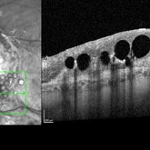
 Retinoschisis with Outer Layer Holes
Jul 18 2025 by Kimberly Wakester
Optomap RGB of an 56-year-old woman with retinoschisis with outer layer holes s/p laser in the left eye. Patient remains stable. Will continue follow up care with dilated exam and optos imaging.
Photographer: Kimberly Wakester, COA, OCT-C
Imaging device: Optos California
Condition/keywords: outer layer hole, retinoschisis
-
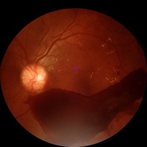 Subhyaloid Hemorrhage
Jul 16 2025 by Paulina Araujo
55-degree central fundus photograph of the left eye (OS) shows a prominent subhyaloid hemorrhage in the inferior posterior pole, displaying a characteristic 'boat-shaped' appearance with well-defined margins and dark red coloration.
Photographer: Paulina D.Araujo Martínez, Asociación para Evitar la Ceguera en México I.A.P., Hospital Dr Luis Sánchez Bulnes.
Condition/keywords: subhyaloid hemorrhage
-
 Large Subhyaloid Hemorrhage
Jul 11 2025 by Jessilla Phou
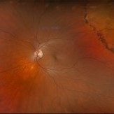
This is a fundus photograph depicting a large subhyaloid hemorrhage in the mid periphery of the left eye. The patient, a 53-year-old female, presented with a sudden onset of floaters, headache, and blurred vision. The image also demonstrates associated optic disc hemorrhage, vitreous hemorrhage, retinal hemorrhage, and venous tortuosity. Despite the extensive workup performed and the severity of the hemorrhage, no underlying cause was determined.
Photographer: Jessilla Phou
Imaging device: Optos California
Condition/keywords: fundus photograph, optic disc hemorrhage, retinal hemorrhage, venous tortuosity, vitreous hemorrhage
-
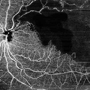 Dark Territories
Jun 27 2025 by Tejaswita Verma
OCT-Angio image of the LE of a 76 year old hypertensive male with history of old superotemporal branch retinal vein occlusion status post 3 anti VEGF injections received in 2012.Vision was 6/9 in left eye. OCTA shows a large CNP area .
Photographer: Dr. Tejaswita Verma
Imaging device: MIRANTE
Condition/keywords: branch retinal vein occlusion (BRVO), CNP areas, OCTA, ST BRVO
-
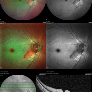 CRAO With Cilio-retinal Sparing-MMI
Jun 25 2025 by Shivankar Sen, MS, FVRS
A 41 year old male came with complaints of Right eye blurring of vision since a day associated with watering and redness. He had no systemic illness, though gave a history of fall from bike 1 month back at the time of which he had blunt force trauma to the right side of the face. BCVA was 3/60, less than N36 in the right eye and 6/6, N6 in the left eye. Right eye had Marcus Gunn Pupil with clear lens, Left eye was within normal limits. IOP was normal; 16 in OD and 18 in OS. Retina evaluation revealed CRAO in the right eye with cilio-retinal artery sparing. Left eye was unremarkable Image Details Left to Right (Top 2 rows) Multicolor Reflectance Image (Blue-green enhanced 55 degree) revealing cilioretinal spared retinal stroma and a characteristic Cherry Red Spot; Green Reflectance showing corresopnding dark gray area with spared perfusion and black spot consistent with Cherry Red Spot on multicolor 2nd Row - 35 degree image (Multicolor Standard Reflectance and Green Reflectance) 3rd Row - SD-OCT revealing acute moderate CRAO findings with Middle retinal layer opacification and prominent middle limiting membrane (p-MLM) sign; Inner retinal layer opacification and prominent retinal pigment epithelium at the fovea with Diminished inner retinal layer stratification
Photographer: Gayathri M S
Imaging device: Heidelberg Spectralis HRA+OCT
Condition/keywords: CRAO with cilioretinal sparing, multicolor, multimodal imaging, OCT biomarkers, reflectance
-
 Berlins Edema - Multimodal Imaging
Jun 25 2025 by Shivankar Sen, MS, FVRS
A 22 year old female came with history of injury to her left eye with a badminton racquet butt cap an hour before presentation On examination, she was found to have right eye 6/6;N6 vision and within normal limits, left eye 6/9;N6 vision, cells1+ in the anterior chamber, brisk pupillary response, no vitreous reaction and sub-clinical berlin's edema at the posterior pole. Multimodal imaging revealed frank boundaries of Berlin's edema more pronounced in the nasal parafoveal region. Figure details Top (Left to Right) Multicolor Reflectance showing bright yellow ring surrounding the perifovea; Blue Reflectance (Black on white contrast) showing corresponding black ring; Green Reflectance showing a characteristic white ring (all pronounced nasally); Bottom (Left-Right) Transverse structural OCT enface image showing white ring consistent with edema OCTA inner layer segmentation from ILM to GCL Transverse corresponding OCTA revealing faint hypo ring within perifoveal capillary bed
Photographer: Gayathri M S
Imaging device: Heidelberg Spectralis HRA+OCT
Condition/keywords: blue reflectance, En Face OCTA, enface imaging, multicolor, oct, reflectance
-
 Berlins
Jun 25 2025 by Shivankar Sen, MS, FVRS
A 22 year old female came with history of injury to her left eye with a badminton racquet butt cap an hour before presentation On examination, she was found to have right eye 6/6;N6 vision and within normal limits, left eye 6/9;N6 vision, cells1+ in the anterior chamber, brisk pupillary response, no vitreous reaction and sub-clinical berlin's edema at the posterior pole. Multimodal imaging revealed frank boundaries of Berlin's edema more pronounced in the nasal parafoveal region. Figure details Top (Left to Right) Multicolor Reflectance showing bright yellow ring surrounding the perifovea; Blue Reflectance (Black on white contrast) showing corresponding black ring; Green Reflectance showing a characteristic white ring (all pronounced nasally); Bottom (Left-Right) Transverse structural OCT enface image showing white ring consistent with edema OCTA inner layer segmentation from ILM to GCL
Photographer: Gayathri M S
Imaging device: Heidelberg Spectralis HRA+OCT
Condition/keywords: blue reflectance, En Face OCTA, multicolor
-
 Active Multi Focal Choroiditis
Jun 21 2025 by Moazzam Parvez
Auto fluorescence image of a 28 year old gentleman with active multifocal choroiditis in his left eye and healed choroiditic patches in the right eye.
Photographer: Moazzam Parvez , Netralayam , Kolkata
Imaging device: Heidelberg Spectralis
Condition/keywords: active, multifocal choroiditis
-
 Subretinal PFO
Jun 18 2025 by Korey Starkey
86-year-old patient had history for retinal detachment surgery x2 and intraocular injections for AMD performed elsewhere. Left eye has PVR developing and subretinal PFO. Due to guarded vision, opting to defer any further treatment at this time.
Photographer: Korey Starkey
Imaging device: Heidelberg
Condition/keywords: AMD, Heidelburg Spectralis, OCT, PFO, PVR, retinal detachment, silicone oil
-
 Uveal Effusion Syndrome
Jun 13 2025 by Brandon I Fram, MD, BS
75 year-old with bilateral inferior serous detachments, right more than left. Scleral window with biopsy showed scleral thickening with stromal deposits of amorphous glycosaminoglycan-like material.
Imaging device: Fluorescein Angiography
Condition/keywords: exudative retinal detachment, idiopathic uveal effusion syndrome, leopard spots, uveal effusion, uveal effusion syndrome
-
 Berlin's Edema
Jun 12 2025 by Shivankar Sen, MS, FVRS
A 22 year old male came with history of sports injury to the right eye with the nose of shuttlecock while playing badminton. On examination, right eye anterior segment shows conjunctival congestion with brisk pupillary reaction and quiet anterior chamber. His best corrected visual acuity was 6/12; N6 in the right eye and 6/6; N6 in the left eye. Retinal examination revealed OD Berlin's Edema, OS within normal limits. Image Description (From Left to Right) Multicolor Reflectance (Blue-Green Enhanced) shows well defined yellowish discoloration Green reflectance and blue reflectance show corresponding whitish discoloration at the area of edema
Photographer: Dr. Shivankar Sen
Imaging device: Heidelberg Spectralis HRA+OCT
Condition/keywords: Shuttlecock Injury
-
 Commotio Retinae
Jun 10 2025 by CUI YUELING
The patient presented 2 hours after sustaining a left eye injury caused by a stick. Visual acuity in the left eye was 0.2 without improvement upon correction, and intraocular pressure measured 15 mmHg. Examination of the anterior segment revealed ciliary conjunctival injection accompanied by patchy subconjunctival hemorrhage. The corneal surface remained smooth, and the anterior chamber was deep with hyphema characterized by blood-tinged aqueous humor predominantly settled inferiorly. The pupil was slightly irregular, approximately 3 mm in diameter, with a superotemporal notch; pupillary light reflex was intact. The lens appeared clear. Fundus examination showed well-defined optic disc margins with normal coloration and a cup-to-disc ratio of 0.2. Retinal arteries and veins were normally distributed with an artery-to-vein ratio of 2:3. At the posterior pole, the foveal reflex exhibited concentric ripple-like changes centered on the fovea, accompanied by localized pigment attenuation and reduced reflex intensity. Irregular reflectivity was noted in the superotemporal and inferotemporal nerve fiber layers.
Photographer: Yueling Cui
Imaging device: Zeiss Clarus 500
Condition/keywords: commotio retinae
 Loading…