Search results (3502 results)
-
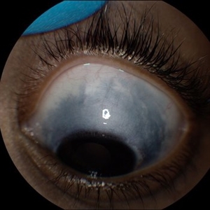 Oculodermal Melanocytosis OS
Jul 5 2024 by Zach Seim
Topcon photograph of a 13 year old with Oculodermal Melanocytosis OS.
Photographer: Zach Seim
Imaging device: Topcon 50DX
Condition/keywords: left, left eye, Oculodermal Melanocytosis, OS, Topcon
-
 “Bull's eye” pattern maculopathy
Mar 14 2023 by Anfisa Ayalon, MD
Left eye fundus autofluorescence image of a 38-year-old female with “Bull's eye” pattern maculopathy. There is no history of medication use associated with retinal toxicity. BCVA LE 20/20-3
Photographer: Danielle Ferguson and Alec Bertoni, University of Pittsburgh Medical Center
Condition/keywords: bull's eye maculopathy, Maculopathy, retina
-
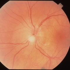
 Acute Retinal Necrosis
Mar 26 2019 by Gary R. Cook, MD, FACS
Left eye of same patient with acute retinal necrosis who developed rhegmatogenous RD seven weeks after presentation.
Imaging device: Topcon VT-50
Condition/keywords: acute retinal necrosis
-
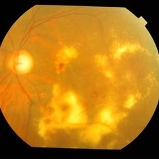
 Acute Toxoplasmosis in AIDS
Apr 8 2019 by Gary R. Cook, MD, FACS
Left eye of a white male with AIDS and an optic neuritis secondary to ocular toxoplasmosis infection. The patient had no pre-existing chorioretinal scars secondary to Toxo. An edematous optic nerve with a focus of active retinitis inferonasally, small surface hemorrhage above it, and surrounding peripapillary edema is visible.
Imaging device: Topcon VT-50
Condition/keywords: AIDS, ocular toxoplasmosis, optic neuritis, toxoplasmosis
-
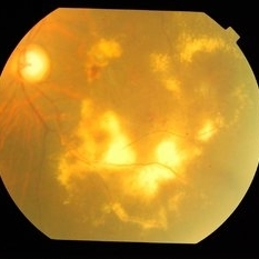
 Adult Coats' Disease
Aug 18 2015 by Mallika Goyal, MD
Left fundus of a 61-year-old non diabetic, non hypertensive lady complaining of vision deterioration for 1 year showed massive hard exudates at the macula. Fluorescein angiography revealed microvascular abnormalities over the posterior pole and temporal midperiphery and extensive capillary non-perfusion over the temporal retinal quadrants. OCT revealed macular edema. Fellow eye fundus and angiogram were normal.
Photographer: Mallika Goyal, MD, Apollo Health City, Jubilee Hills, Hyderabad
Condition/keywords: Coats' disease
-
 Adult Coats' Disease
Aug 18 2015 by Mallika Goyal, MD
Left fundus of a 61-year-old non diabetic, non hypertensive lady complaining of vision deterioration for 1 year showing massive hard exudates at the macula. Fluorescein angiography revealed microvascular abnormalities over the posterior pole and temporal midperiphery and extensive capillary non-perfusion over the temporal retinal quadrants. OCT revealed macular edema. Fellow eye fundus and angiogram were normal.
Photographer: Mallika Goyal, MD, Apollo Health City, Jubilee Hills, Hyderabad
Condition/keywords: Coats' disease
-
 Adult Coats' Disease
Aug 18 2015 by Mallika Goyal, MD
Left fundus of a 61-year-old non diabetic, non hypertensive lady complaining of vision deterioration for 1 year showed massive hard exudates at the macula. Fluorescein angiography revealed microvascular abnormalities over the posterior pole and temporal midperiphery and extensive capillary non-perfusion over the temporal retinal quadrants. OCT revealed macular edema. Fellow eye fundus and angiogram were normal.
Photographer: Mallika Goyal, MD, Apollo Health City, Jubilee Hills, Hyderabad
Condition/keywords: Coats' disease
-
 Adult Foveomacular Dystrophy
Aug 21 2025 by Aditya S Kelkar, MS, FRCS, FASRS,FRCOphth
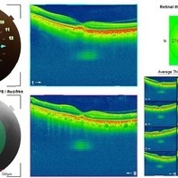
Left eye OCT of a 56 year old female with complaints of gradual painless blurring of vision, aggravating on near work.
Photographer: Dr. Muskan Mangal
Condition/keywords: adult foveomacular dystrophy, OCT
-
---thumb.jpg/image-square;max$300,300.ImageHandler) Adult Vitelliform Dystrophy
Adult Vitelliform Dystrophy
Feb 13 2013 by From the Collections of Thomas M. Aaberg, MD and Thomas M. Aaberg Jr., MD
Left eye
Condition/keywords: left eye, vitelliform macular dystrophy
-
 Adult Vitelliform Macular Dystrophy
Jun 25 2024 by Tejaswita Verma
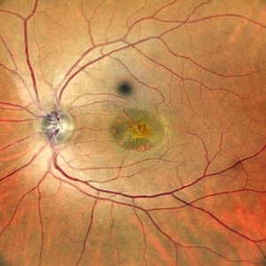
Left eye fundus photograph of an elderly 62 year old hypertensive female showing elevated lesion at macula s/o adult vitelliform macular dystrophy, misdiagnosed as long standing CSR elsewhere.
Photographer: DR. TEJASWITA VERMA
Imaging device: MIRANTE
Condition/keywords: Adult vitelliform macular dystrophy
-
---thumb.JPG/image-square;max$300,300.ImageHandler) Advanced AMD with extensive scarring and bleed
Advanced AMD with extensive scarring and bleed
Dec 8 2013 by Mallika Goyal, MD
Left eye of 70-year-old gentleman with bilateral extensive scarring and bleeding from advanced AMD 6 months after anti-VEGF Therapy.
Photographer: Mallika Goyal, MD, Apollo Health City, Hyderabad, India
-
---thumb.JPG/image-square;max$300,300.ImageHandler) Advanced AMD With Extensive Scarring and Bleeding
Advanced AMD With Extensive Scarring and Bleeding
Dec 8 2013 by Mallika Goyal, MD
Left eye of 70-year-old gentleman with bilateral extensive scarring and bleed from advanced AMD 6 months after anti-VEGF Therapy.
Photographer: Mallika Goyal, MD, Apollo Health City, Hyderabad, India
-
---thumb.JPG/image-square;max$300,300.ImageHandler) Advanced AMD With Extensive Scarring and Bleeding
Advanced AMD With Extensive Scarring and Bleeding
Dec 8 2013 by Mallika Goyal, MD
Left eye of an elderly male with bilateral advanced AMD with extensive scarring and bleed 6 months after anti-VEGF therapy.
Photographer: Mallika Goyal, MD, Apollo Health City, Hyderabad, India
-
---thumb.JPG/image-square;max$300,300.ImageHandler) Advanced AMD With Extensive Scarring and Bleeding
Advanced AMD With Extensive Scarring and Bleeding
Dec 8 2013 by Mallika Goyal, MD
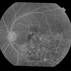
Left eye fluorescein angiogram of an elderly male with bilateral advanced AMD with extensive scarring and bleeding 6 months after anti-VEGF therapy.
Photographer: Mallika Goyal, MD, Apollo Health City, Hyderabad, India
-
---thumb.JPG/image-square;max$300,300.ImageHandler) Advanced AMD With Extensive Scarring and Bleeding
Advanced AMD With Extensive Scarring and Bleeding
Dec 8 2013 by Mallika Goyal, MD
Left eye fluorescein angiogram of an elderly male with bilateral advanced AMD with extensive scarring and bleeding 6 months after anti-VEGF therapy.
Photographer: Mallika Goyal, MD, Apollo Health City, Hyderabad, India
 Loading…