 Initializing download.
-
 By Tejaswita Verma
Retina Foundation hospital(Ahmedabad)
Co-author(s): Dr. Manish Nagpal,RETINA FOUNDATION AHMEDABAD - Uploaded on Jun 27, 2025.
- Last modified by Joshua Friedman on Jun 30, 2025.
- Rating
- Appears in
- Miscellaneous
- Condition/keywords
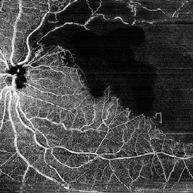
- OCTA, branch retinal vein occlusion (BRVO), ST BRVO, CNP areas
- Photographer
- Dr. Tejaswita Verma
- Imaging device
-
Optical coherence tomography system
MIRANTE - Description
- OCT-Angio image of the LE of a 76 year old hypertensive male with history of old superotemporal branch retinal vein occlusion status post 3 anti VEGF injections received in 2012.Vision was 6/9 in left eye. OCTA shows a large CNP area .


 Caused due Branch Retinal Vein Occlusion (BRVO)")
 after Anti VEGF Treatment")
")


