Search results (52 results)
-
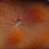
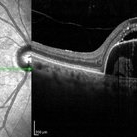
 Macular Mount Everest
Aug 8 2025 by Anand Temkar
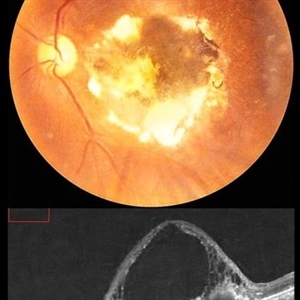
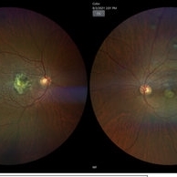
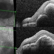
A 75 yrs old male came with the chief complains of DOV in LE since past 20 yrs. His BCVA in RE was 6/9 and in LE, it was CF 1 meter. His IOP was 13 mm of Hg in RE and 15 mm of Hg in LE. Patient is a k/c/o DM type 2 since past 20 yrs and is on regular medication. Patient is a k/c/o solitary kidney. Patient gives h/o ( LE ) Intravitreal injection Avastin 3 times 13 yrs ago i/c/o CNVM. In the LE color photo we can see the scarred CNVM along with altered foveal contour. LE OCT also shows cystic spaces with large elevation and scarring.
Photographer: Dr.Anand Temkar- Vasan Eye Hospital, Tiruchirapalli
Condition/keywords: CNVM, macular edema, scarred cnvm
-
 Central Retinal Vein Occlusion With Waldenstroms macroglobulinemia
Jun 18 2025 by Korey Starkey
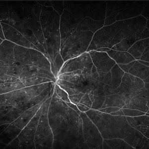
64-year-old patient presents with CRVO with secondary macular edema in both eyes. Venous beading present in 2/4 quadrants OU. Patient diagnosed with Waldenstroms macroglobulinemia, found on SPEP and bone marrow biopsy. Treatment recommended of anti-vegF intravitreal injections OU.
Photographer: Korey Starkey
Imaging device: Optos
Condition/keywords: attenuated vessels, central retinal vein occlusion (CRVO), CRVO, FA early phase, FLUORESCEIN ANGIOGRAPHY, macular edema, Optomap, OPTOS CALIFORNIA, severe NPDR, venous beading, Waldenstroms macroglobulinemia
-
 Superior Rhegmatogenous Retinal Detachment (RRD) in the Right Eye, With a Retinal Tear Located Between the 1 and 2 O'clock Positions
Apr 4 2025 by Cesar Orlando Oviedo Vera
A 45-year-old male patient presented with a sudden onset of decreased visual acuity in the right eye, with a 24-hour progression. Upon examination, Image 1 revealed a superior rhegmatogenous retinal detachment in the right eye, with a retinal tear located between the 1 and 2 o'clock positions. Image 2: Pneumatic retinopexy by intravitreal injection of Sulfur Hexafluoride gas (SF6) at the time of diagnosis with subsequent application of 532 nm laser around the retinal tear.
Photographer: Cesar Orlando Oviedo Vera, Hospital Militar de Especialidades Oftalmológicas
Imaging device: Optos
Condition/keywords: Pneumatic Retinopexy, Retinal tear, Rhegmatogenous retinal detachment, SF6, Superior rhegmatogenous retinal detachment
-
 Pneumatic Retinopexy by Intravitreal Injection of Sulfur Hexafluoride Gas (SF6) at the Time of Diagnosis With Subsequent Application of 532 Nm Laser Around the Retinal Tear
Apr 4 2025 by Cesar Orlando Oviedo Vera
A 45-year-old male patient presented with a sudden onset of decreased visual acuity in the right eye, with a 24-hour progression. Upon examination, Image 1 revealed a superior rhegmatogenous retinal detachment in the right eye, with a retinal tear located between the 1 and 2 o'clock positions. Image 2: Pneumatic retinopexy by intravitreal injection of Sulfur Hexafluoride gas (SF6) at the time of diagnosis with subsequent application of 532 nm laser around the retinal tear.
Photographer: Cesar Orlando Oviedo Vera, Hospital Militar de Especialidades Oftalmológicas
Imaging device: Optos
Condition/keywords: Pneumatic Retinopexy, Retinal tear, Rhegmatogenous retinal detachment, SF6, Superior rhegmatogenous retinal detachment
-
 Ozurdex Implant
Mar 12 2025 by Virginia Gebhart
65 year old female 4 weeks s/p intravitreal Ozurdex implant for diabetic macular edema. Significant improvement of edema and SRF
Photographer: Virginia Gebhart, Retina Consultants of Carolina
Imaging device: Optos California
Condition/keywords: diabetic macular edema, DME, intravitreal implant, intravitreal injection, ozurdex, Ozurdex implant
-
 Central Retinal Vein Occlusion With Macular Edema
Feb 27 2025 by Kimberly Wakester
Optomap RGB image of a 34-year-old woman with central retinal vein occlusion with macular edema in the left eye. Patient has had a fairly acute onset central retinal vein occlusion in her left eye with dense superior IRH and macular edema. Modest ischemic changes are seen on exam and fundus photos. Patient was educated on the etiology of CRVOs and the relationship to systemic risk factors. Recommended hypercoagulable work-up with her PCP and bloodwork was ordered. Treatment with intravitreal injections was recommended to reduce the macular edema. Patient is to continue monthly follow ups with repeat OCT.
Photographer: Kimberly Wakester, COA
Imaging device: Optos California
Condition/keywords: CRVO with macular edema
-
 Central Retinal Vein Occlusion with Macular Edema
Jan 29 2025 by Kimberly Wakester
Fundus photograph of a 62-year-old man with central retinal vein occlusion with macular edema and a new PVD with an operculated retinal tear in the left eye. Laser to retinal tear was completed. Patient will return in 2-3 weeks for follow up exam with possible intravitreal injection for the CRVO with edema and to follow up on the operculated retinal tear s/p retinal tear laser.
Photographer: Kimberly Wakester, COA
Imaging device: Optos California
Condition/keywords: central retinal vein occlusion (CRVO), operculated tear, PVD
-
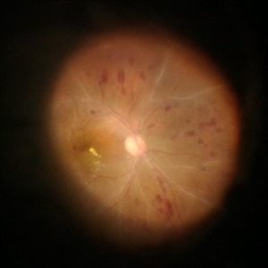
 Atypical Tubercular Occlusive Peripheral Retinal Vasculitis
Jun 21 2024 by Tejaswita Verma
Follow up right eye fundus photograph of a 27 year old male with vision 6/12 , diagnosed with systemic tuberculosis(mediastinal lymphadenopathy on chest CT) on oral steroids, and started on ATT .We can see a parafoveal sub-ILM hemorrhage with vascular sheathing and hemorrhages in inferior and temporal quadrants . The patient was advised anti-VEGF intravitreal injection, later sectoral laser after resolution of inflammation
Photographer: DR. TEJASWITA VERMA
Imaging device: MIRANTE
Condition/keywords: obliterative peripheral vasculitis, ocular tuberculosis
-
 Pigment Epithelium Detachment, Secondary to AMD
Mar 17 2023 by Ceara Donovan
Optical coherence tomography of a 76 year old woman with a Pigment Epithelium Detachment, Secondary to AMD affecting her right eye. Patient had no significant response to Avastin, Eylea, Lucentis 0.5, or Vabysmo and was switched to Beovu. Following Beovu intravitreal injection her edema improved on OCT. Patient's vision was sc20/200+1 at the time the image was taken.
Photographer: Ceara Donovan
Imaging device: Heidelberg Spectralis
Condition/keywords: exudative age-related macular degeneration, heidelberg spectralis, macular degeneration, optical coherence tomography (OCT), pigment epithelial detachment (PED), Sub-retinal fluid
-
 Inflammatory pupillary membrane in patient with endophthalmitis
Jan 28 2023 by Kingston Rodolfo Ureña-Wong, MD, Opht, MSc
Anterior segment photography of a 54-year-old woman with post phacoemulsification endophthalmitis. She did not improve after first intravitreal antibiotics injection and develop an inflammatory pupillary membrane. After two vitrectomies, and a complete three intravitreal injections scheme, we decided to remove the intraocular lens and capsules.
Photographer: Marco Antonio Rubio-Atonal,UNAM, Asociación para evitar la ceguera en México
Imaging device: Zeiss Clarus 700
Condition/keywords: endophthalmitis, pupillary membranes
-
 A rare case of a 45-year-old male with choroidal neovascular membrane in Familial Dominant Drusen (Doyne Honeycomb Drusen) in both eyes treated with intravitreal injections.
Nov 30 2022 by SHRADDHA ASHOK CHANDORKAR, DNB DO FVRS
A 45-year-old man presented with diminution of vision in both eyes with metamorphopsia, which was painless and gradually progressive in nature. BCVA at presentation were 6/40 and 6/36 for the right and left eye respectively. Anterior segment examination of both eyes was unremarkable. IOP were within normal limits. Fundus examination showed bilateral numerous yellowish white round closely spaced lesions extending radially from the vascular arcades till the periphery associated with an elevated grayish macular choroidal neovascular membrane (CNV) with multiple drusen in the macular area and posterior pole. Impression was Familial Dominant Drusen (Doyne Honeycomb Drusen) associated with CNVM, both eyes. Color fundus photograph and autofluorescence showed Familial Dominant Drusen with CNVM. Subsequently , the patient underwent periodic intravitreal injections of Ranibizumab in both eyes under guarded visual prognosis, for which he tolerated well.
Photographer: NATIONAL INSTITUTE OF OPHTHALMOLOGY, PUNE
Imaging device: ZEISS CLARUS
Condition/keywords: choroidal neovascular membrane (CNVM), Doyne's Honeycomb, FAMILIAL DOMINANT DRUSEN, IMIM (Online Mendelian Inheritance in Man), intravitreal injection, Malattia Leventinese
-
 Methotrexate Bubble following Intravitreal Injection for PVR
Sep 21 2022 by Zach Seim
Ultra-widefield fundus photograph of an 81 year old female with a Methotrexate bubble following an Intravitreal Injection for Proliferative Vitreoretinopathy. Patient has been presenting to the office for two week interval Methotrexate injections in her left eye. The image was taken prior to her eighth injection which revealed a residual Methotrexate bubble in her inferior retinal image. This patient was seeing "lots" of floaters, as well as having visual acuity of cc20/400 cc20/200 PH.
Photographer: Zach Seim
Imaging device: OPTOS California
Condition/keywords: bubble, fundus photograph, fundus photography, intravitreal injection, left eye, methotrexate, nasal retina, Optos, proliferative vitreoretinopathy (PVR), pseudocolor, ultra-wide field imaging
-
 Submacular Hemorrhage after C3F8 Pneumatic Displacement
Jul 5 2021 by Fang Helen Mi
The patient underwent urgent intravitreal injection of recombinant tissue plasminogen activator and pneumatic displacement with 100% C3F8 gas. The following day, the sub-macular haemorrhage was largely displaced away from the fovea inferiorly and nasally, with the gas bubble seen superiorly.
Imaging device: Clarus Camera
Condition/keywords: gas pneumatic displacement, polypoidal choroidal vasculopathy (PCV), submacular hemorrhage
-
 Post Injection Endophthalmitis
Nov 21 2020 by Suhwan Lee, MD
Stillcut image of eye with post-intravitreal injection endophthalmitis during emergency vitrectomy. The patient received intravitreal injection for neovascular age-related macular degeneration 2 days ago. Image that was taken after anterior chamber irrigation and core vitrectomy showed multiple retinal hemorrhage and whitening of retinal vessels.
Photographer: Suhwan Lee, MD
Imaging device: Stillcut image of surgical video (Lumera 700 )
Condition/keywords: endophthalmitis
-
 cRORA
cRORA
Aug 5 2020 by Dhaivat Shah
A 54-year-old healthy male presented to us with a decreased vision in right eye since past 8 years. The patient gave a history of bleed in right eye before 8 years for which some intravitreal injection was given; post which there no major visual improvement. No details or documentation was available regarding the same. His BCVA in the right eye was 5/60. Fundus examination revealed a sharply demarcated hypopigmented patch over the macula with mild posterior excavation suggestive of macular scar. OCT image shows foveal thinning with loss of Retinal pigment epithelium and outer retinal layers (RORA). There are 2 types of RORAs, complete and incomplete. Complete RORA and incomplete RORA are entities defined by various imaging modalities describing atrophy of the retinal pigment epithelial and the outer retinal layers. OCT imaging defines incomplete RORA (iRORA) as a region of signal hyper transmission into the choroid and a corresponding zone of attenuation ordisruption of the RPE (<250um) and evidence of overlying photoreceptor degeneration (<250um). There should not be any RPE tear associated with it. OCT imaging describes complete RORA (cRORA) based on 4 inclusion criteria. These include, area of hypertransmission of more than 250um, zone of attenuation or disruption of the RPE of more than 250um in diameter, evidence of overlying photoreceptor degeneration and absence of scrolled RPE or other signs of an RPE tear. Other modalities used to define these include fundus autoflourescence(FAF), near infrared reflectance(NIR) and color fundus photograph(CFP). On CFP, it shows a sharply demarcated hypopigmented of >250um size with better visibility of choroidal vessels. FAF shows a hypo autoflourescent patch with sharply demarcated borders of size >250um, the colour of which is similar to that of the optic nerve head or retinal blood vessels excluding any pigmentation or artefact. On NIR, it shows a hyperreflective area with sharply demarcated borders of >250um size excluding any artefact. RORA can be seen in conditions like geographical atrophy in ARMD, central areolar choroidal dystrophy, atrophy secondary to anti-VEGF treatment. References: 1. Sadda SR, Guymer R, Holz FG, et al. Consensus Definition for Atrophy Associated with Age-Related Macular Degeneration on OCT: Classification of Atrophy Report 3 [published correction appears in Ophthalmology. 2019 Jan;126(1):177]. Ophthalmology. 2018;125(4):537-548. 2. Guymer RH, Rosenfeld PJ, Curcio CA, et al. Incomplete Retinal Pigment Epithelial and Outer Retinal Atrophy in Age-Related Macular Degeneration: Classification of Atrophy Meeting Report 4. Ophthalmology. 2020;127(3):394-409. 3. Eng VA, Rayess N, Nguyen HV, Leng T. Complete RPE and outer retinal atrophy in patients receiving anti-VEGF treatment for neovascular age-related macular degeneration. PLoS One. 2020;15(5):e0232353.
Photographer: Miss Anjum Zafar Khan
Imaging device: Choithram Netralaya
Condition/keywords: macular scar, outer retina, retinal pigment epithelium
-
 Intravitreal Air Bubbles after anti-VEGF Injection
Jul 24 2020 by Darin R. Goldman, MD
Widefield fundus photograph of a 56-year-old male patient immediately following intravitreal anti-VEGF injection. Two air bubbles are visible within the mid-vitreous cavity.
Photographer: Alberto Velez, Retina Group of Florida
Imaging device: Zeiss Clarus
Condition/keywords: anti-VEGF, intravitreal, intravitreal injection
-
 Satisfactory Long-Term Visual Outcome after Radiation Retinopathy Treatment
Jun 5 2020 by Sophia El Hamichi, MD
A 71-year-old female treated with plaque brachytherapy for choroidal melanoma in her left eye 10 years ago. She subsequently developed radiation retinopathy OS for which she was regularly receiving intravitreal injections of bevacizumab. The result is a stable visual acuity at 20/30. The patient continues to be regularly monitored.
Photographer: Belinda Rodriguez
Condition/keywords: bevacizumab, optical coherence tomography (OCT), radiation retinopathy
-
 Satisfactory Long-Term Visual Outcome after Radiation Retinopathy Treatment
May 26 2020 by Sophia El Hamichi, MD
A 71-year-old female treated with plaque brachytherapy for choroidal melanoma in her left eye 10 years ago. She subsequently developed radiation retinopathy OS for which she was regularly receiving intravitreal injections of bevacizumab. The result is a stable visual acuity at 20/30. The patient continues to be regularly monitored.
Photographer: Belinda Rodriguez, Murray Ocular Oncology and Retina, Miami
Condition/keywords: intravitreal bevacizumab, optical coherence tomography (OCT), radiation retinopathy
-
 Radiation Retinopathy Post Brachytherapy Plaque Treating Choroidal Melanoma
May 26 2020 by Sophia El Hamichi, MD
A 71-year-old female treated with plaque brachytherapy for choroidal melanoma in her left eye 10 years ago. She subsequently developed radiation retinopathy OS for which she was regularly receiving intravitreal injections of bevacizumab. The result is a stable visual acuity at 20/30. The patient continues to be regularly monitored.
Photographer: Belinda Rodriguez, Murray Ocular Oncology and Retina, Miami
Condition/keywords: I-125 brachytherapy, melanoma, radiation retinopathy
-
 Management of Submacular Hemorrhage
Mar 22 2020 by Anfisa Ayalon, MD
Submacular hemorrhage in age-related macular degeneration. Image A: SD-OCT before the surgery. Note widespread submacular hemorrhage with subretinal fluid and RPE detachment. Image B: SD-OCT after the surgery (PPV+ERM and ILM peeling+Submacular injection of t-PA, bevacizumab and filtered air+ Intravitreal injection of 20% SF6 ). Note remaining RPE detachment with no OCT signs of submacular hemorrhage. The visual acuity improved after the surgery from HM to 1/60.
Photographer: Anfisa Ayalon, MD., Meir Medical Center, Kfar Saba, Israel.
Condition/keywords: age-related macular degeneration (AMD), optical coherence tomography (OCT), submacular hemorrhage
-
 Management of Submacular Hemorrhage
Mar 22 2020 by Anfisa Ayalon, MD
Intraoperative images were taken during the management of submacular hemorrhage in age-related macular degeneration. The goal of the surgery was the physical displacement of SMH out of the fovea using expansile gas. The image from the left was done during ILM peeling. Note the massive collection of subretinal blood. The image from the right was done after submacular injection of t-PA, bevacizumab and filtered air. Intravitreal injection of 20% SF6 completed the surgery. The visual acuity improved after the surgery from HM to 1/60.
Photographer: Anfisa Ayalon, MD., Meir Medical Center, Kfar Saba, Israel.
Condition/keywords: spontaneous submacular hemorrhage, submacular hemorrhage, vitreomacular surgery, wet age-related macular degeneration (wet AMD)
-
 Choroidal Osteoma
Jan 22 2020 by Gregg T. Kokame, MD, MMM, FASRS
45-year-old female, with choroidal osteoma. Silicone oil droplet in the periphery, after intravitreal injection.
Photographer: Jaclyn Pisano, Retina Consultants of Hawaii
Imaging device: Clarus 700
Condition/keywords: choroidal osteoma, silicone oil
-
 Choroidal Osteoma
Jan 22 2020 by Gregg T. Kokame, MD, MMM, FASRS
45-year-old female, with choroidal osteoma. silicone oil droplet in the periphery, after intravitreal injection.
Photographer: Jaclyn Pisano
Imaging device: Clarus 700
Condition/keywords: choroidal osteoma, intravitreal injection, silicone oil
-
 Retinopathy of Prematurity Stage 4A Treated with Laser and Bevacizumab Intravitreal Injection
Jan 6 2020 by Sophia El Hamichi, MD
A 41-week-old girl, with a gestational age of 23 weeks and 4 days presenting with stage 4 A of ROP OU. She was treated with laser and intravitreal injections of bevacizumab.
Photographer: Abby Orcutt-Hayes, Murray Ocular Oncology and Retina
Condition/keywords: intravitreal bevacizumab, laser photocoagulation, retinopathy of prematurity stage 4a
-
 Montage Photo: Retinopathy of Prematurity Stage 4A Treated with Laser and Bevacizumab Intravitreal Injection
Jan 6 2020 by Sophia El Hamichi, MD
A 41-week-old girl, with a gestational age of 23 weeks and 4 days presenting with stage 4 A of ROP OU. She was treated with laser and intravitreal injections of bevacizumab.
Photographer: Abby Orcutt-Hayes, Murray Ocular Oncology and Retina
Condition/keywords: intravitreal bevacizumab, laser photocoagulation, montage, retinopathy of prematurity stage 4a
 Loading…