 Initializing download.
-
 By Anfisa Ayalon, MD
McGill University
Co-author(s): Alexander Rubowitz, MD.,Chen Shtayer, MD., Meir Medical Center, Kfar Saba, Israel. - Uploaded on Mar 22, 2020.
- Last modified by Caroline Bozell on Mar 24, 2020.
- Rating
- Appears in
- Miscellaneous
- Condition/keywords
- optical coherence tomography (OCT), submacular hemorrhage, age-related macular degeneration (AMD)
- Photographer
- Anfisa Ayalon, MD., Meir Medical Center, Kfar Saba, Israel.
- Imaging device
- Optical coherence tomography system
- Description
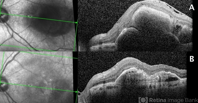
- Submacular hemorrhage in age-related macular degeneration. Image A: SD-OCT before the surgery. Note widespread submacular hemorrhage with subretinal fluid and RPE detachment. Image B: SD-OCT after the surgery (PPV+ERM and ILM peeling+Submacular injection of t-PA, bevacizumab and filtered air+ Intravitreal injection of 20% SF6 ). Note remaining RPE detachment with no OCT signs of submacular hemorrhage. The visual acuity improved after the surgery from HM to 1/60.

")


")
")
")


