 Initializing download.
-
 By Anfisa Ayalon, MD
McGill University
Co-author(s): Alexander Rubowitz, MD., Chen Shtayer, MD., Meir Medical Center, Kfar Saba, Israel. - Uploaded on Mar 22, 2020.
- Last modified by Caroline Bozell on Mar 24, 2020.
- Rating
- Appears in
- Miscellaneous
- Condition/keywords
- submacular hemorrhage, wet age-related macular degeneration (wet AMD), vitreomacular surgery, spontaneous submacular hemorrhage
- Photographer
- Anfisa Ayalon, MD., Meir Medical Center, Kfar Saba, Israel.
- Imaging device
- Fundus camera
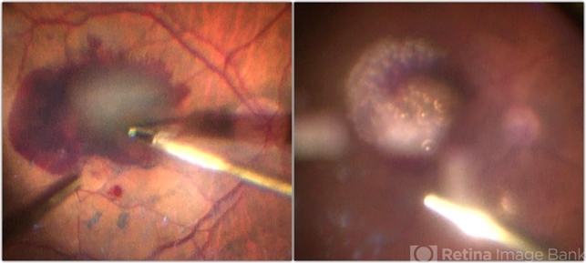
- Description
- Intraoperative images were taken during the management of submacular hemorrhage in age-related macular degeneration. The goal of the surgery was the physical displacement of SMH out of the fovea using expansile gas. The image from the left was done during ILM peeling. Note the massive collection of subretinal blood. The image from the right was done after submacular injection of t-PA, bevacizumab and filtered air. Intravitreal injection of 20% SF6 completed the surgery. The visual acuity improved after the surgery from HM to 1/60.


---thumb.JPG/image-square;max$79,0.ImageHandler "spontaneous submacular hemorrhage")

---thumb.JPG/image-square;max$79,0.ImageHandler "spontaneous submacular hemorrhage")
---thumb.JPG/image-square;max$79,0.ImageHandler "Spontaneous Submacular Hemorrhages")
---thumb.JPG/image-square;max$79,0.ImageHandler "spontaneous submacular hemorrhage")


