Search results (44 results)
-
 Emulsified Silicon Oil in Macular Hole- Hyperoleon in Hole
Mar 8 2025 by PUJA NEGI
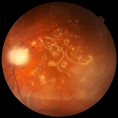
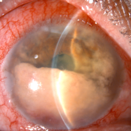
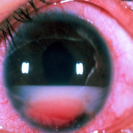
Fundus photograph showing emulsified silicon oil bubbles in macular hole, hyperoleon in hole.
Photographer: Dr Vaidehi Jethwa
Condition/keywords: Inverse hypopyon, macular hole
-
 Best Disease
Apr 24 2024 by Marcelo Zas, MD PhD
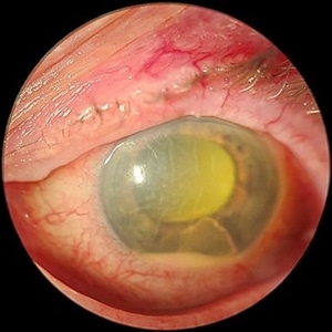
Best vitelliform macular dystrophy (BVMD) or Best disease. Is the most common autosomal dominant macular dystrophy. It involves the retinal pigment epithelium (RPE), and leads to a characteristic bilateral yellow “egg-yolk” appearance of the macula as you can see in this image. Essentially, BVMD is considered to have 6 clinical stages: Previtelliform, Vitelliform, Pseudohypopyon, Vitelleruptive, Atrophic and Choroidal neovascularization. As the disease progresses, patients may experience a slow, bilateral decrease in visual acuity, central scotoma, or metamorphopsia. With secondary CNV, visual decline can be rapid, however.
Photographer: Luciano Scorsetti MD
Condition/keywords: Macular Dystrophy
-
 Acute Endophthalmitis
Nov 14 2023 by Virginia Gebhart
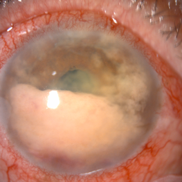
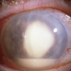
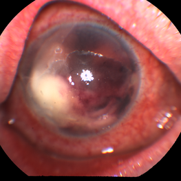
85 year old female with acute endophthalmitis 14 days s/p IVEylea injection. 3+ injection and descemets folds, contracted fibron, 3mm Hypopyon, and 3+ cell. No view posteriorly, vision CF. Visual prognosis unknown at this time
Photographer: Virginia Gebhart
Condition/keywords: endophthalmitis
-
 Retinoblastoma Pseudohypopyon
Dec 10 2022 by Jordan D Deaner, MD
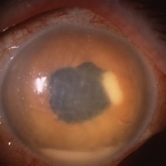
6-year-old female referred for left eye pain with elevated intraocular pressure, neovascularization of the iris, and a hypopyon concerned for uveitis eventually diagnosed with pseudohypopyon secondary to diffuse infiltrating retinoblastoma.
Condition/keywords: Retinoblastoma Pseudohypopyon
-
 Secondary intraocular lymphoma
Apr 11 2022 by Aniruddha K Agarwal, MD
A 65-year-old male underlying nasopharyngeal non-Hodgkin’s lymphoma presented with pseudo-hypopyon and infiltration of iris from tumor on his right eye. Aqueous tap showed atypical lymphocytes.
Photographer: Kessara Pathanapitoon MD, PhD Department of Ophthalmology, Faculty of Medicine Chiang Mai University, Chiang Mai, Thailand
Condition/keywords: lymphoma, masquerade syndrome, secondary iridocyclitis (noninfectious)
-
 Secondary intraocular lymphoma
Apr 11 2022 by Aniruddha K Agarwal, MD
A 65 year-old male underlying nasopharygeal non-Hodgkin’s lymhoma presented with pseudohypopyon and infiltration of iris from tumor on his right eye. Aqueous tap showed atypical lymphocytes.
Photographer: Kessara Pathanapitoon MD, PhD Department of Ophthalmology, Faculty of Medicine Chiang Mai University, Chiang Mai, Thailand
Condition/keywords: lymphoma, masquerade syndrome, secondary iridocyclitis (noninfectious)
-
 Adult-onset vitelliform dystrophy
Mar 28 2022 by T. P . VIGNESH, MBBS,MS
Fundus photo of right eye of a 45 year male patient with pseudohypopyon stage of Adult-onset vitelliform dystrophy .
Photographer: Bharathi Singaravel
Imaging device: Zeiss Clarus
Condition/keywords: Adult-onset vitelliform dystrophy
-
 Mushroom-shaped Choroidal Melanoma
May 18 2020 by McGill University Health Centre
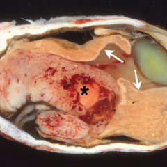
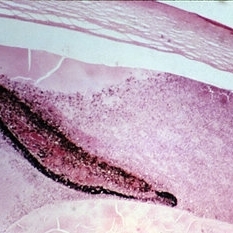
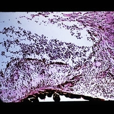
The enucleation specimen in (A) shows a diffuse melanoma infiltrating the choroid and ciliary body. In the center, a large area of necrosis and hemorrhage is present (*) and the retina is infiltrated (arrows). Note hypopyon in the anterior chamber and the cataractous lens.
Condition/keywords: enucleation, mushroom-shaped
-
 Toxoplasmic Uveitis
May 18 2020 by McGill University Health Centre
This enucleation specimen shows multiple irregular chorioretinal scars, surrounded by hyperplastic retinal pigment epithelium (arrowheads). Some adherent membranes are present in the vitreous cavity (arrow). The lens has a total cataract and the anterior chamber is filled with whitish material that corresponds to hypopyon.
Condition/keywords: toxoplasmosis uveitis, uveitis
-
 Lens-Induced Uveitis
May 18 2020 by McGill University Health Centre
In lens-induced uveitis, lens protein in the anterior chamber causes a zonal granulomatous response, which occurs usually 1 day to 3 weeks after capsule rupture. This may be associated with sympathetic ophthalmia. This enucleation specimen shows an indented cornea, accompanied by complete hypopyon occupying the anterior chamber with intense vitreitis. Note that the translucent vitreous has become whitish, and the lens surface is irregular with decoloration of the peripheral areas (arrows).
Condition/keywords: uveitis
-
 Large B Cell Lymphoma of the Retina
Dec 13 2019 by McGill University Health Centre
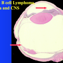
65-year-old female with the clinical diagnosis of bilateral uveitis of unknown ethiology. Histopathology of a large B cell lymphoma in the anterior chamber, causing pseudohypopyon.
Photographer: Miguel N. Burnier, McGill University Health Center-McGill University Ocular Pathology & Translational Research Laboratory
Imaging device: Zeiss
Condition/keywords: large b cell lymphoma of the retina, masquerade syndrome, pseudohypopyon
-
 Large B Cell Lymphoma of the Retina
Dec 13 2019 by McGill University Health Centre
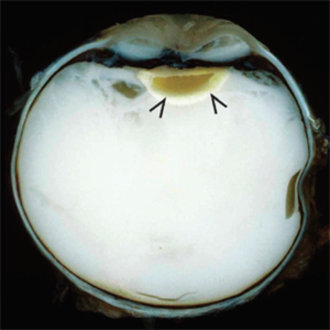
65-year-old female with the clinical diagnosis of bilateral uveitis of unknown etiology. Histopathology of the enucleated specimen showing large neoplastic cells in the anterior chamber, representing pseudohypopyon in a case of masquerade syndrome. The retina shows areas of necrosis with neoplastic large B cells.
Photographer: Miguel N. Burnier, McGill University Health Center-McGill University Ocular Pathology & Translational Research Laboratory
Imaging device: Zeiss
Condition/keywords: large b cell lymphoma, large b cell lymphoma of the retina, masquerade syndrome, pesudohypopyon
-
 Large B Cell Lymphoma of the Retina
Dec 13 2019 by McGill University Health Centre
65-year-old female with the clinical diagnosis of bilateral uveitis of unknown ethiology. The clinical picture shows a large pseudohypopyon, consistent with large B cell lymphoma of the retina, vitreous and CNS.
Photographer: Miguel N. Burnier, McGill University Health Center-McGill University Ocular Pathology & Translational Research Laboratory
Condition/keywords: large b cell lymphoma, pseudohypopyon, retina, uveitis, vitreous
-
 Reverse Reverse Hypopyon
Jul 15 2019 by Anfisa Ayalon, MD
Slit-lamp photograph of a pseudo-hypopyon of perfluorocarbon in the eye of a young patient who had undergone repair of rhegmatogenous retinal detachment years prior. Note the emulsified appearance.
Photographer: Anfisa Ayalon, MD. Meir Medical Center, Kfar Saba, Israel.
Condition/keywords: pars plana vitrectomy (PPV), perfluorocarbon fluid, retained perfluorocarbon
-
 Fungal Endophthalmitis
Jun 4 2019 by Gary R. Cook, MD, FACS
Fungal (Candida) endophthalmitis with hypopyon.
Condition/keywords: candida endophthalmitis, endophthalmitis, fungal endophthalmitis, hypopyon
-
 Endophthalmitis
Apr 1 2019 by Gary R. Cook, MD, FACS
Klebsiella pneumonia endophthalmitis with hypopyon post cataract surgery.
Condition/keywords: endogenous endophthalmitis, endophthalmitis, klebsiella pneumoniae
-
 Slide 13-17
Mar 4 2019 by Lancaster Course in Ophthalmology
Extension of retinoblastoma to involve the iris and anterior chamber with a pseudophypopyon present ( x25).
Condition/keywords: pseudohypopyon, retinoblastoma
-
 Slide 3-9
Feb 20 2019 by Lancaster Course in Ophthalmology
Low-power view of eye with Candida albicans endophthalmitis ( x12). Note the hypopyon and exudate in the anterior chamber and the infiltration of the iris and ciliary body with inflammatory cells.
Condition/keywords: ciliary, endophthalmitis, exudate, hypopyon, iris
-
 Slide 2-1
Feb 19 2019 by Lancaster Course in Ophthalmology
Hypopyon in a suppurative endophthalmitis following cataract surgery.
Condition/keywords: cataract, endophthalmitis, hypopyon
-
 Slide 1-5
Feb 19 2019 by Lancaster Course in Ophthalmology
Pus (hypopyon) in the angle of the anterior chamber, secondary to a bactecorneal ulcer. (H&E stain)
Condition/keywords: bactecorneal ulcer, hypopyon, pus
-
 Intraocular Eyelash
Jan 2 2019 by John S. King, MD
34-year-old white male injured while taking apart wooden pallets with a hammer in each hand and no protective eye-wear; he did not notice what hit his eye; just said his eye hurt and teared for four days before calling an eye doctor; his vision was 20/400 sc and IOP 6; the anterior chamber was deep with minimal cell and no hypopyon; and conjunctival/scleral laceration was present near the lateral rectus insersion; the vitreous was quiet; in the temporal portion of the fundus and full thickness laceration was seen with surrounding hemorrhage and what appeared to be an eyelash vs other (possibly a staple, from the wooden pallet). During surgery 2 eyelashes were pulled from the area of the laceration; the lateral rectus muscle was disorganized; after primary closure, a ppv was performed, the object in the picture was removed with forcepts; laser partial afx and gas; antibiotics injected at 1/2 dose.
Photographer: Maisee Yang
Imaging device: Optos CA
Condition/keywords: intraocular foreign body, ruptured globe
-
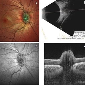 ONH-Drusen
-
 Inverse Hypopyon
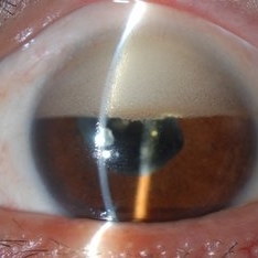
Mar 4 2018 by Yoshihiro Yonekawa, MD, FASRS
Slit lamp photograph of a 40-year-old man with previous retinal detachment surgery with silicone oil tamponade, presenting with an inverse hypopyon from emulsified silicone oil.
Photographer: Steven A Bennett, COA, CRA
Imaging device: Nikon D200 / Topcon Slit lamp
Condition/keywords: hypopyon, silicone oil
-
 Endophthalmitis
Feb 22 2018 by Nichole Lewis
Endophthalmitis
Photographer: Nichole Lewis
Condition/keywords: endophthalmitis, hypopyon
 Loading…