Search results (44 results)
-
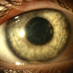
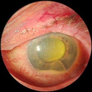
 Corneal Ulcer
Sep 14 2017 by Theodore Leng, MD, MS, FASRS
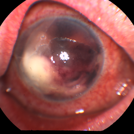
Corneal ulcer with hypopyon
Condition/keywords: central corneal ulcer, hypopyon
-
 Endophthalmitis
Feb 22 2018 by Nichole Lewis
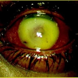
Endophthalmitis
Photographer: Nichole Lewis
Condition/keywords: endophthalmitis, hypopyon
-
 Endophthalmitis Complicating an Intraocular Foreign Body
Jul 12 2014 by Philip J. Polkinghorne, MD
An 8-year-old child presented with endophthalmitis following an injury to the eye. At the time of surgery a retained foreign body was found, latter identified as a fragment from a pencil.
Photographer: Philip Polkinghorne
Condition/keywords: endophthalmitis, hypopyon, intraocular foreign body
-
 Fungal Endophthalmitis
Jun 4 2019 by Gary R. Cook, MD, FACS
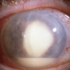
Fungal (Candida) endophthalmitis with hypopyon.
Condition/keywords: candida endophthalmitis, endophthalmitis, fungal endophthalmitis, hypopyon
-
 HLA-B27 Associated Uveitis
Jun 4 2014 by Henry J. Kaplan, MD
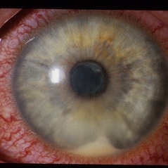
Severe anterior uveitis with fibrinous reaction and hypopyon formation related to HLA-B27. Notice the membrane on the lens surface.
Condition/keywords: acute anterior uveitis, HLA-B27, hypopyon
-
 Hypopyon
Oct 9 2012 by Jeffrey G. Gross, MD, FASRS
Hypopyon, broken synechia, HLA, B 27 syndrome.
Condition/keywords: B 27 syndrome, broken synechia, HLA-B27, hypopyon
-
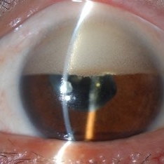 Inverse Hypopyon
Mar 4 2018 by Yoshihiro Yonekawa, MD, FASRS
Slit lamp photograph of a 40-year-old man with previous retinal detachment surgery with silicone oil tamponade, presenting with an inverse hypopyon from emulsified silicone oil.
Photographer: Steven A Bennett, COA, CRA
Imaging device: Nikon D200 / Topcon Slit lamp
Condition/keywords: hypopyon, silicone oil
-
 Inverted Hypopyon - Silicon Oil Complication
Feb 12 2015 by H. Michael Lambert, MD
Silicon oil emulsification, inverted hypopyon.
Condition/keywords: hypopyon, silicone oil
-
 Inverted Hypopyon - Silicon Oil Complication
Feb 12 2015 by H. Michael Lambert, MD
Silicon oil emulsification, inverted hypopyon.
Condition/keywords: hypopyon, silicone oil
-
 Inverted Hypopyon - Silicon Oil Complication
Feb 12 2015 by H. Michael Lambert, MD
Silicon oil emulsification, inverted hypopyon.
Condition/keywords: hypopyon, silicone oil
-
 Inverted Hypopyon - Silicon Oil Complication
Feb 12 2015 by H. Michael Lambert, MD
Silicon oil emulsification, inverted hypopyon.
Condition/keywords: hypopyon, silicone oil
-
 ONH-Drusen
-
 Removing a Hypopyon From the Anterior Segment
Jul 12 2014 by Philip J. Polkinghorne, MD
An intra-operative photograph demonstrating the technique of removing a hypopyon from the anterior segment.
Photographer: Philip Polkinghorne
Condition/keywords: endophthalmitis, fibrin, hypopyon
-
 Slide 1-5
Feb 19 2019 by Lancaster Course in Ophthalmology
Pus (hypopyon) in the angle of the anterior chamber, secondary to a bactecorneal ulcer. (H&E stain)
Condition/keywords: bactecorneal ulcer, hypopyon, pus
-
 Slide 2-1
Feb 19 2019 by Lancaster Course in Ophthalmology
Hypopyon in a suppurative endophthalmitis following cataract surgery.
Condition/keywords: cataract, endophthalmitis, hypopyon
-
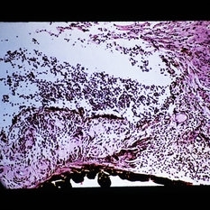
 Slide 3-9
Feb 20 2019 by Lancaster Course in Ophthalmology
Low-power view of eye with Candida albicans endophthalmitis ( x12). Note the hypopyon and exudate in the anterior chamber and the infiltration of the iris and ciliary body with inflammatory cells.
Condition/keywords: ciliary, endophthalmitis, exudate, hypopyon, iris
-
 Subhyaloid Hemorrhage, Hypopyon
Oct 19 2012 by Larry Halperin, MD
Subhyaloid hemorrhage, hypopyon
Condition/keywords: hypopyon, subhyaloid hemorrhage
-
 Acute Endophthalmitis
Nov 14 2023 by Virginia Gebhart
85 year old female with acute endophthalmitis 14 days s/p IVEylea injection. 3+ injection and descemets folds, contracted fibron, 3mm Hypopyon, and 3+ cell. No view posteriorly, vision CF. Visual prognosis unknown at this time
Photographer: Virginia Gebhart
Condition/keywords: endophthalmitis
-
 Adult-onset vitelliform dystrophy
Mar 28 2022 by T. P . VIGNESH, MBBS,MS
Fundus photo of right eye of a 45 year male patient with pseudohypopyon stage of Adult-onset vitelliform dystrophy .
Photographer: Bharathi Singaravel
Imaging device: Zeiss Clarus
Condition/keywords: Adult-onset vitelliform dystrophy
-
 Best Disease
Jun 4 2014 by Henry J. Kaplan, MD
Best disease, pseduohypopyon formation.
Condition/keywords: Best disease, pseudohypopyon
-
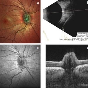
 Best Disease
Sep 28 2016 by Maciej Czepita
Color fundus image and fundus autofluorescence image of a 41-year-old male patient with Best disease (pseudohypopyon stage).
Photographer: Maciej Czepita, Pomeranian Medical University, Szczecin, Poland
Imaging device: Heidelberg Spectralis HRA+OCT
Condition/keywords: Best disease
-
 Best Disease
Apr 24 2024 by Marcelo Zas, MD PhD
Best vitelliform macular dystrophy (BVMD) or Best disease. Is the most common autosomal dominant macular dystrophy. It involves the retinal pigment epithelium (RPE), and leads to a characteristic bilateral yellow “egg-yolk” appearance of the macula as you can see in this image. Essentially, BVMD is considered to have 6 clinical stages: Previtelliform, Vitelliform, Pseudohypopyon, Vitelleruptive, Atrophic and Choroidal neovascularization. As the disease progresses, patients may experience a slow, bilateral decrease in visual acuity, central scotoma, or metamorphopsia. With secondary CNV, visual decline can be rapid, however.
Photographer: Luciano Scorsetti MD
Condition/keywords: Macular Dystrophy
-
 Best's disease
Apr 4 2013 by Jerald A. Bovino, MD
some pseudohypopion, probably Best's
Condition/keywords: Best disease, pseudohypopyon
-
 Bests Disease Multi Focal OS
Oct 9 2012 by Alan D. Letson, MD
Multifocal vitelliform lesions and "pseudohypopyon"
Photographer: Beverly Radcliffe
Condition/keywords: pseudohypopyon, vitelliform lesion
-
---thumb.jpg/image-square;max$300,300.ImageHandler) Bilateral Hypopyon
Bilateral Hypopyon
Jan 2 2014 by David Callanan, MD
Bilateral hypopyon, 11-year-old male.
Condition/keywords: bilateral hypopyn
 Loading…