Search results (44 results)
-
 Vitelliform Macular Dystrophy or Best Disease
Dec 16 2016 by Young Hee Yoon, MD, PhD
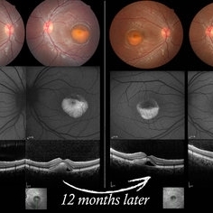
Bilateral fundus photographs and autofluorescence images of 15-year-old girl who was diagnosed as vitelliform macular dystrophy or Best disease. Vitelliform macular lesion showed morphologic change during one year.
Photographer: Hyejin Jo, Sunghyun Kim, Heoni Hong, Minjung Chae, Mihwa Shin, Asan medical center, Seoul
Imaging device: Topcon TRC-500X fundus camera, Heidelberg HRA 2 autofluorescence, Heldelberg Spectralis OCT
Condition/keywords: Best disease, pseudohypopyon, scrambled-egg, vitelliform macular dystrophy
-
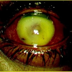
 HLA-B27 Associated Uveitis
Jun 4 2014 by Henry J. Kaplan, MD
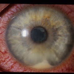
Severe anterior uveitis with fibrinous reaction and hypopyon formation related to HLA-B27. Notice the membrane on the lens surface.
Condition/keywords: acute anterior uveitis, HLA-B27, hypopyon
-
 Inverted Hypopyon - Silicon Oil Complication
Feb 12 2015 by H. Michael Lambert, MD
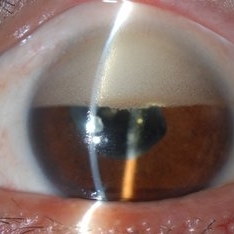
Silicon oil emulsification, inverted hypopyon.
Condition/keywords: hypopyon, silicone oil
-
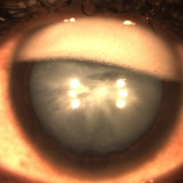
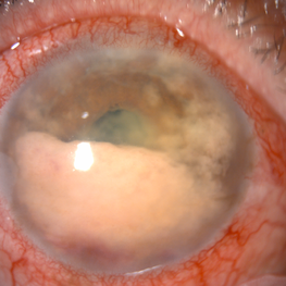
 Hypopyon
Oct 9 2012 by Jeffrey G. Gross, MD, FASRS
Hypopyon, broken synechia, HLA, B 27 syndrome.
Condition/keywords: B 27 syndrome, broken synechia, HLA-B27, hypopyon
-
 Inverted Hypopyon - Silicon Oil Complication
Feb 12 2015 by H. Michael Lambert, MD
Silicon oil emulsification, inverted hypopyon.
Condition/keywords: hypopyon, silicone oil
-
 Inverse Hypopyon
Mar 4 2018 by Yoshihiro Yonekawa, MD, FASRS
Slit lamp photograph of a 40-year-old man with previous retinal detachment surgery with silicone oil tamponade, presenting with an inverse hypopyon from emulsified silicone oil.
Photographer: Steven A Bennett, COA, CRA
Imaging device: Nikon D200 / Topcon Slit lamp
Condition/keywords: hypopyon, silicone oil
-
 Subhyaloid Hemorrhage, Hypopyon
Oct 19 2012 by Larry Halperin, MD
Subhyaloid hemorrhage, hypopyon
Condition/keywords: hypopyon, subhyaloid hemorrhage
-
 Best Disease
Jun 4 2014 by Henry J. Kaplan, MD
Best disease, pseduohypopyon formation.
Condition/keywords: Best disease, pseudohypopyon
-
 IOFB-Endophthalmitis Slide 2
Oct 22 2012 by Ronald C. Gentile, MD
Anterior segment examination revealed a self sealing peripheral corneal wound at the 7:30 position with iris defect. There was a small hypopyon layering in the inferior angle. CT scan revealed a small intra-ocular foreign body.
Photographer: The New York Eye & Ear Infirmary Department of Medical Imaging
Condition/keywords: intraocular foreign body
-
 Best Disease
Sep 28 2016 by Maciej Czepita
Color fundus image and fundus autofluorescence image of a 41-year-old male patient with Best disease (pseudohypopyon stage).
Photographer: Maciej Czepita, Pomeranian Medical University, Szczecin, Poland
Imaging device: Heidelberg Spectralis HRA+OCT
Condition/keywords: Best disease
-
 Bests Disease Multi Focal OS
Oct 9 2012 by Alan D. Letson, MD
Multifocal vitelliform lesions and "pseudohypopyon"
Photographer: Beverly Radcliffe
Condition/keywords: pseudohypopyon, vitelliform lesion
-
 Inverse hypopyon-Emulcified silicon Oil
Apr 24 2015 by Mehul A Shah
Patient had history of vitrectomy with silicon oil injection before 6 years.
Photographer: Mehul Shah
Imaging device: Zeiss FF450plus
Condition/keywords: Inverse hypopyon, silicone oil
-
 Removing a Hypopyon From the Anterior Segment
Jul 12 2014 by Philip J. Polkinghorne, MD
An intra-operative photograph demonstrating the technique of removing a hypopyon from the anterior segment.
Photographer: Philip Polkinghorne
Condition/keywords: endophthalmitis, fibrin, hypopyon
-
 Corneal Ulcer
Sep 14 2017 by Theodore Leng, MD, MS, FASRS
Corneal ulcer with hypopyon
Condition/keywords: central corneal ulcer, hypopyon
-
 Inverted Hypopyon - Silicon Oil Complication
Feb 12 2015 by H. Michael Lambert, MD
Silicon oil emulsification, inverted hypopyon.
Condition/keywords: hypopyon, silicone oil
-
 Inverted Hypopyon - Silicon Oil Complication
Feb 12 2015 by H. Michael Lambert, MD
Silicon oil emulsification, inverted hypopyon.
Condition/keywords: hypopyon, silicone oil
-
 Endophthalmitis Complicating an Intraocular Foreign Body
Jul 12 2014 by Philip J. Polkinghorne, MD
An 8-year-old child presented with endophthalmitis following an injury to the eye. At the time of surgery a retained foreign body was found, latter identified as a fragment from a pencil.
Photographer: Philip Polkinghorne
Condition/keywords: endophthalmitis, hypopyon, intraocular foreign body
-
 Bleb-related Endophthalmitis Slide 2
Oct 22 2012 by Ronald C. Gentile, MD
Anterior chamber has a hypopyon with fibrin.
Photographer: The New York Eye & Ear Infirmary Department of Medical Imaging
Condition/keywords: Bleb-related endophthalmitis
-
 Endophthalmitis
Feb 22 2018 by Nichole Lewis
Endophthalmitis
Photographer: Nichole Lewis
Condition/keywords: endophthalmitis, hypopyon
-
 Reverse Reverse Hypopyon
Jul 15 2019 by Anfisa Ayalon, MD
Slit-lamp photograph of a pseudo-hypopyon of perfluorocarbon in the eye of a young patient who had undergone repair of rhegmatogenous retinal detachment years prior. Note the emulsified appearance.
Photographer: Anfisa Ayalon, MD. Meir Medical Center, Kfar Saba, Israel.
Condition/keywords: pars plana vitrectomy (PPV), perfluorocarbon fluid, retained perfluorocarbon
-
 Secondary intraocular lymphoma
Apr 11 2022 by Aniruddha K Agarwal, MD
A 65-year-old male underlying nasopharyngeal non-Hodgkin’s lymphoma presented with pseudo-hypopyon and infiltration of iris from tumor on his right eye. Aqueous tap showed atypical lymphocytes.
Photographer: Kessara Pathanapitoon MD, PhD Department of Ophthalmology, Faculty of Medicine Chiang Mai University, Chiang Mai, Thailand
Condition/keywords: lymphoma, masquerade syndrome, secondary iridocyclitis (noninfectious)
-
 Best's disease
Apr 4 2013 by Jerald A. Bovino, MD
some pseudohypopion, probably Best's
Condition/keywords: Best disease, pseudohypopyon
-
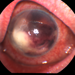
 Fungal Endophthalmitis
Jun 4 2019 by Gary R. Cook, MD, FACS
Fungal (Candida) endophthalmitis with hypopyon.
Condition/keywords: candida endophthalmitis, endophthalmitis, fungal endophthalmitis, hypopyon
-
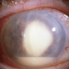
---thumb.jpg/image-square;max$300,300.ImageHandler) Bilateral Hypopyon
Bilateral Hypopyon
Jan 2 2014 by David Callanan, MD
Bilateral hypopyon, 11-year-old male.
Condition/keywords: bilateral hypopyn
-
 Intraocular Eyelash
Jan 2 2019 by John S. King, MD
34-year-old white male injured while taking apart wooden pallets with a hammer in each hand and no protective eye-wear; he did not notice what hit his eye; just said his eye hurt and teared for four days before calling an eye doctor; his vision was 20/400 sc and IOP 6; the anterior chamber was deep with minimal cell and no hypopyon; and conjunctival/scleral laceration was present near the lateral rectus insersion; the vitreous was quiet; in the temporal portion of the fundus and full thickness laceration was seen with surrounding hemorrhage and what appeared to be an eyelash vs other (possibly a staple, from the wooden pallet). During surgery 2 eyelashes were pulled from the area of the laceration; the lateral rectus muscle was disorganized; after primary closure, a ppv was performed, the object in the picture was removed with forcepts; laser partial afx and gas; antibiotics injected at 1/2 dose.
Photographer: Maisee Yang
Imaging device: Optos CA
Condition/keywords: intraocular foreign body, ruptured globe
 Loading…