Search results (189 results)
-
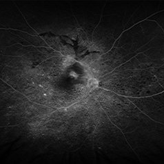
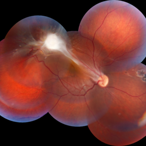
 The Retinal Tempest: Toxocara's Trail
Aug 31 2025 by Giriraj Vibhute
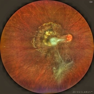
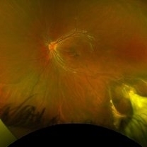
In this striking image, a central white granuloma spirals outward from the optic disc, surrounded by fibrous traction bands and scarring—the telltale markings of intraocular toxocara lesion. The retina is ravaged with proliferative vitreoretinal membranes and peripheral pigmentary changes, starkly illustrating the chronic inflammation and vision-threatening complications caused by Toxocara canis.
Photographer: Dr Giriraj Vibhute, MM Joshi eye institute, Hubli, India.
Condition/keywords: dragged disc, fibrous proliferation, Toxocara, toxocara canis
-
 Acute Retinal Necrosis (ARN)
Jul 3 2025 by Heitor Nogueira
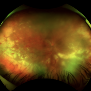
Fundus photograph of an 63-year-old woman who reported unilateral visual acuity loss for 10 days associated with ocular pain. He presented conjunctival hyperemia with temporal and nasal nodular scleritis, anterior chamber reaction 2+/4+, Koeppe nodules, granulomatous PKs, vitreitis 2+/4+, multiple areas of vasculitis in the arcades and periphery, associated with hemorrhages and necrotizing retinitis in the temporal, inferior and nasal periphery. Positive serology for Herpes Virus
Photographer: Heitor Nogueira, Penido Burnier Institute, Campinas, São Paulo, Brazil
Imaging device: Optos Daytona
Condition/keywords: ARN complications, Herpes, progressive outer retinal necrosis (PORN), Uveitis
-
 Acute Retinal Necrosis
Jul 3 2025 by Heitor Nogueira
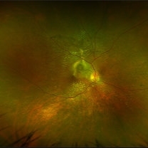
Fundus photograph of an 53-year-old woman with patient who reported unilateral visual acuity loss for 10 days associated with ocular pain. She presented conjunctival hyperemia with temporal and nasal nodular scleritis, anterior chamber reaction 2+/4+, Koeppe nodules, granulomatous PKs, vitritis 2+/4+, multiple areas of vasculitis in arcades and periphery, associated with hemorrhages and necrotizing retinitis in temporal, inferior and nasal periphery. patient who reported unilateral visual acuity loss for 10 days associated with ocular pain. He presented conjunctival hyperemia with temporal and nasal nodular scleritis, anterior chamber reaction 2+/4+, Koeppe nodules, granulomatous PKs, vitreitis 2+/4+, multiple areas of vasculitis in the arcades and periphery, associated with hemorrhages and necrotizing retinitis in the temporal, inferior and nasal periphery. Positive serology for Herpes Virus.
Photographer: Heitor Nogueira, Penido Burnier Institute and CHOV, Campinas, São Paulo, Brazil
Imaging device: Optos Daytona
Condition/keywords: ARN complications, Herpes, progressive outer retinal necrosis (PORN)
-
 The Headlight in the Fog
Jun 17 2025 by Thirumalesh Mochi Basavaraj, MD
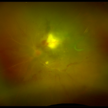
37 year old male with sudden onset diminution of visual acuity has a large retinochoridal granuloma along the superotemporal arcade and a few with satellite lesions more temporal to it, there was extensive Occlusoive vasculitis (both arterioles and veins )being involved with Vitrities.
Photographer: Vivekanand ,Narayana nethralaya
Imaging device: Daytona
Condition/keywords: acute toxoplasmosis, retinochoroiditis
-
 VKH Pseudotumor – Chronic Subretinal Fibrosis
May 11 2025 by Felipe Murati
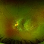
Ultra-widefield fundus image from a 36-year-old woman with chronic VKH syndrome showing a pseudotumor-like subretinal fibrotic lesion in the right eye. The lesion developed after multiple relapses and remained stable over a 1-year follow-up with immunosuppressive treatment including prednisone, mycophenolate mofetil, and adalimumab. No active choroiditis or exudative detachment was observed. Multimodal imaging was essential for disease monitoring.
Photographer: Felipe A. Murati, MD, University of Arizona
Imaging device: Optos California ultra-widefield retinal imaging system, single-capture, color fundus modality.
Condition/keywords: adalimumab, chronic inflammation, granulomatous uveitis, OCT, Optos ultra-widefield imaging, pseudotumor, subretinal fibrosis, VKH, Vogt-Koyanagi-Harada
-
 VKH Pseudotumor – Fluorescein Angiography
May 11 2025 by Felipe Murati
Fluorescein angiography image from a 36-year-old woman with chronic Vogt-Koyanagi-Harada (VKH) syndrome showing a pseudotumor-like lesion with late-phase staining and no active leakage. The image highlights subretinal fibrosis in the right eye, stable under long-term immunosuppressive therapy with mycophenolate mofetil and adalimumab. No signs of active choroiditis are present, confirming a quiescent phase.
Photographer: Felipe A. Murati, MD, University of Arizona
Imaging device: Optos California, fluorescein angiography modality
Condition/keywords: choroiditis, Fluorescein angiography, granulomatous uveitis, Optos FA, pseudotumor, subretinal fibrosis, VKH, Vogt-Koyanagi-Harada
-
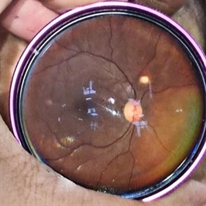
 Optic Disc Granuloma
May 7 2025 by Aayesha - Khanum, MBBS. D.N.B
A 45-year-old male presented with diminished vision for one month. His Mantoux test was negative, but as steroids worsened the condition, quantiferon TB was advised and it returned positive. He was started on anti-tuberculosis treatment (ATT). Oral steroids were reintroduced after one week of ATT. Optic disc granulomas can arise from direct invasion of the optic nerve or may represent hypersensitivity reaction to tuberculous antigens. The pathogenesis involves infiltration of immune cells, leading to formation of a granulomatous structure that disrupts normal architecture and function of the optic disc. Steroids with ATT facilitated regression of granulomatous lesion.
Photographer: Ms. Krishna Jeyanthi
Imaging device: Zeiss Clarus 500
Condition/keywords: Tuberculosis
-
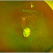
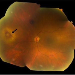
 Toxocara Granuloma
Apr 18 2025 by Chellarani Kumarasamy, MD
10 year old girl referred for starbismus and red free image showed well demarcted inactive lesion.
Condition/keywords: toxocara granuloma
-
 Central Posterior Granuloma
Apr 18 2025 by Chellarani Kumarasamy, MD
Fundus photo showed central posterior granuloma on the optic nerve
Condition/keywords: toxocara granuloma
-
 Tuberculoma
Sep 23 2024 by DR Rohit Gupta
Fundus photograph of a 26 year old female suffering from pulmonary tuberculosis, on fundus examination a tuberculoma seen in her left eye.
Photographer: DR Rohit gupta
Imaging device: Samsung S21
Condition/keywords: tubercular choroidal granuloma
-
 Ocular Toxocariasis
Jul 4 2024 by Brandon I Fram, MD
4 yo with toxocariasis-related peripheral granuloma with adhesion to the macula and macular subretinal fibrosis. Positive Toxocara titers.
Condition/keywords: toxocara canis, toxocara granuloma, toxocariasis
-
 Multimodal Imaging for Differentiating Unilateral Pseudo Optic Disc Swelling(Buried Drusen) From True Optic Disc Swelling
Feb 7 2024 by Fawwaz F Al Mamoori, MD, Medical Retina Consultant
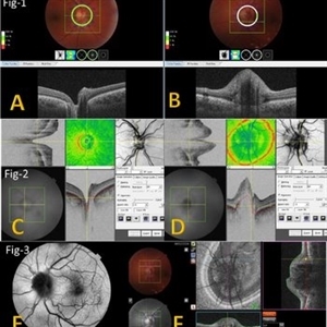
27-year-old male, medically free, presented with left unilateral optic disc swelling. BCVA=1.0(OU), color vision, and contrast sensitivity were normal (OU)with no RAPD in the left eye. Swept Source OCT: showed elevated left optic disc with hyporeflective mass (Fig-1 B). Enface OCT: Showed left peripapillary multiple ovoid mass lesions(drusen) (Fig-2 d, Fig3 F). FAF: of the left eye showed superonasal hyper autofluorescent drusenoid lesions)(Fig3 E). Orbital MRI with contrast was requested to exclude any compressive lesions like tumors(menigioma)or inflammatory lesions like granuloma(sarcoid granuloma). orbital MRI result was normal.
Photographer: Hana.S.Owais
Imaging device: TRITON(TOPCON,Swept Source OCT)
Condition/keywords: fundus autofluorescence (FAF), multimodal imaging, OCT EN FACE, optic disc drusen, optic disc edema, swept source
-
 Multimodal Imaging for Differentiating Unilateral Pseudo Optic Disc Swelling(Buried Drusen) From True Optic Disc Swelling
Feb 7 2024 by Fawwaz F Al Mamoori, MD, Medical Retina Consultant
A 27-year-old male patient, medically free, presented with unilateral left optic disc swelling. BCVA=1.0(OU), color vision, and contrast sensitivity were normal (OU) with no RAPD in the left eye. SS-OCT: showed left optic disc elevation with hyporeflective mass lesion (Fig-1 B). Enface OCT: showed left peripapillary hyperreflective ovoid mass lesions(Fig-2 D, Fig-3 F), FAF: showed left superonasal hyperautofluorescent drusenoid lesions. Orbital MRI with contrast was requested to exclude any optic nerve compressive lesions like (tumors: like mengioma or inflammatory lesions like granuloma (sarcoidosis). the result of orbital MRI was normal.
Photographer: Hana.S.Owais
Imaging device: TRITON(TOPCON,Swept Source OCT)
Condition/keywords: fundus autofluorescence (FAF), multimodal imaging, OCT EN FACE, optic disc drusen, optic disc edema
-
 Fibrotic granuloma vs. Pseudoduplication of the Optic Disc
Nov 29 2023 by Virginia Gebhart
74 year-old female with presumed fibrotic granuloma. Previously diagnosed as pseudoduplication of the optic disc by general ophthalmologist. OCT showed elevation in the RPE, more consistent with granuloma. Pt has been aware for many years, asymptomatic. Will observe.
Photographer: Virginia Gebhart
Imaging device: Topcon 50DX
Condition/keywords: fibrotic scar, granuloma, Pseudoduplication of optic disc
-
 Choroidal Granuloma
Aug 6 2023 by AMIT NENE
Fundus photograph of 27 year old female with choroidal granuloma and disc edema treated with IVMP and oral steroids resulting in complete melt of granuloma at follow-up
Photographer: Gaurav Kamble, Isha Netralaya, Thane
Imaging device: Optos imaging
Condition/keywords: choroidal granuloma, optic disc edema
-
 Choroidal granuloma
Jul 30 2023 by Dr.Sheetal Divate
A 23 year old female came with chief complaints of blurring of vision in left eye since 20 days. Fundus examination disc odema with choroidal granuloma.
Photographer: Dr.Sheetal Divate
Imaging device: Optos Advance
Condition/keywords: choroidal granuloma, Choroidal Lesion
-
 Tubercular Granuloma/Choroidal Granuloma
Jul 17 2023 by Thirumalesh Mochi Basavaraj, MD
Composite Optos+SSOCT image Tubercular granuloma
Photographer: Puttaswamy N K ,Narayana Nethralaya, Bangalore
Condition/keywords: composite image OCT and widefiled image, tubercular choroidal granuloma
-
 Choroidal Granuloma
Jul 17 2023 by Thirumalesh Mochi Basavaraj, MD
Wide filed image of a tubercular choroidal granuloma
Photographer: Puttaswamy N K , Narayana Nethralaya, Bangalore
Condition/keywords: choroidal tuberculoma, tubercular choroidal granuloma
-
 Sarcoidosis
Mar 27 2023 by Joshua Friedman
Fundus photograph of a 70-year-old woman with presumed sarcoidosis and sarcoid-associated choroidal granuloma.
Photographer: Christopher D Conrady, MD, PhD
Condition/keywords: sarcoid granuloma
-
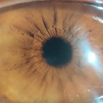 Koeppe Iris nodules
Apr 14 2022 by Divya Jain
Slit Lamp photograph of a 33 year old woman with first episode of acute granulomatous anterior uveitis showing mutton fat KP'S and Koeppe's nodules.
Photographer: Divya Jain
Condition/keywords: acute anterior uveitis
-
 Acute Anterior Uveitis
Apr 14 2022 by Divya Jain
Anterior Segment Slit Lamp photograph of a 33 year old woman with first episode of acute granulomatous anterior uveitis showing circumcorneal congestion, mutton fat KP'S, 3+ cells, 2+ flare and Koeppe's nodules at pupillary margin.
Photographer: Divya Jain
Condition/keywords: acute anterior uveitis
-
 TB Granuloma
Nov 14 2021 by Maxwell J Wingelaar, MD
A tubercular choroidal granuloma in a patient with 4-drug resistant TB.
Condition/keywords: tubercular choroidal granuloma
-
 Peripheral Toxocariasis Granuloma with Macular Traction
Jul 6 2021 by Lucas Zago Ribeiro, MD
15-year-old male patient with vision loss for the last 4 years, presenting peripheral toxocariasis granuloma, and macular traction.
Photographer: Lucas Zago Ribeiro, Federal University of São Paulo (UNIFESP)
Imaging device: Zeiss Visucam 524
Condition/keywords: granuloma, toxocariasis
-
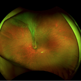 Ocular Toxocariasis with Peripheral Granuloma
Apr 24 2021 by Alexandre Grandinetti, MD, PhD
8-year-old boy with a retinal fold secondary to peripheral toxocara canis granuloma localized on the superior retina.
Photographer: Corina Skrzek
Imaging device: Optos California
Condition/keywords: toxocariasis
-
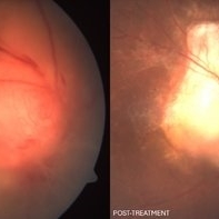
 Bartonella Posterior Granulomatous Mass with Exudative Detachment Post 2 Months
Sep 26 2020 by Swati Agarwal-Sinha, MD, FASRS
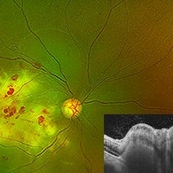
Color fundus photo of the left eye two months post-treatment showing resolved exudative detachment, fibrosed granuloma at the optic disc, tractional detachment at the posterior pole and extensive diffuse subretinal exudates extending all around up to the retinal periphery. No retinal vascular changes are noted.
Photographer: Harry Rosa, University of Florida
Condition/keywords: Bartonella bacteria, exudative detachment
 Loading…