 Initializing download.
-
 By Swati Agarwal-Sinha, MD, FASRS
Northeast Wisconsin Retina Associates
Co-author(s): Swati Agarwal-Sinha - Uploaded on Sep 26, 2020.
- Last modified by Caroline Bozell on Sep 29, 2020.
- Rating
- Appears in
- Miscellaneous
- Condition/keywords
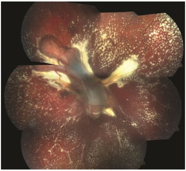
- Bartonella bacteria, exudative detachment
- Photographer
- Harry Rosa, University of Florida
- Imaging device
- Fundus camera
- Description
- Color fundus photo of the left eye two months post-treatment showing resolved exudative detachment, fibrosed granuloma at the optic disc, tractional detachment at the posterior pole and extensive diffuse subretinal exudates extending all around up to the retinal periphery. No retinal vascular changes are noted.


---thumb.jpg/image-square;max$79,0.ImageHandler "Subretinal Hemorrhage With Exudative Detachment")
---thumb.jpg/image-square;max$79,0.ImageHandler "Subretinal Hemorrhage With Exudative Detachment")
---thumb.jpg/image-square;max$79,0.ImageHandler "Subretinal Hemorrhage With Exudative Detachment")
---thumb.jpg/image-square;max$79,0.ImageHandler "Subretinal Hemorrhage With Exudative Detachment")


