Search results (189 results)
-
 Toxocara Granuloma
Feb 25 2013 by Henry J. Kaplan, MD
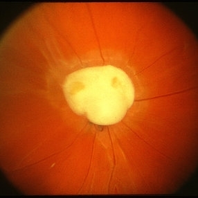
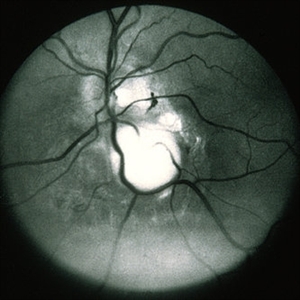
Toxocara granuloma of the optic nerve head.
Condition/keywords: ocular toxoplasmosis, toxocara granuloma, toxocariasis
-
 Acute Retinal Necrosis (ARN)
Jul 3 2025 by Heitor Nogueira
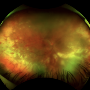
Fundus photograph of an 63-year-old woman who reported unilateral visual acuity loss for 10 days associated with ocular pain. He presented conjunctival hyperemia with temporal and nasal nodular scleritis, anterior chamber reaction 2+/4+, Koeppe nodules, granulomatous PKs, vitreitis 2+/4+, multiple areas of vasculitis in the arcades and periphery, associated with hemorrhages and necrotizing retinitis in the temporal, inferior and nasal periphery. Positive serology for Herpes Virus
Photographer: Heitor Nogueira, Penido Burnier Institute, Campinas, São Paulo, Brazil
Imaging device: Optos Daytona
Condition/keywords: ARN complications, Herpes, progressive outer retinal necrosis (PORN), Uveitis
-
 Choroidal Granuloma
Apr 7 2017 by Manish Nagpal, MD, FRCS (UK), FASRS
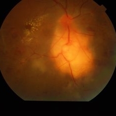
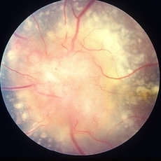
Colour photo of a case of peripapillary choroidal granuloma presenting with exudation and hemorrhages.
Photographer: pooja barot
Condition/keywords: choroid, granuloma, inflammation
-
 Choroidal Granuloma
Apr 7 2017 by Manish Nagpal, MD, FRCS (UK), FASRS
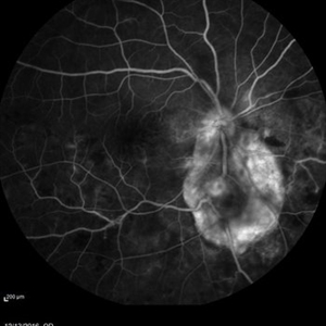
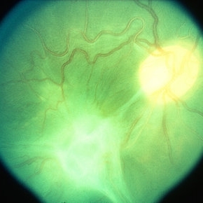
Fluorescein angiography of a case of peripapillary choroidal granuloma presenting with exudation and hemorrhages.
Photographer: Pooja Barot
Condition/keywords: choroid, granuloma, inflammation
-
---thumb.JPG/image-square;max$300,300.ImageHandler) Tubercular choroidal granuloma
Tubercular choroidal granuloma
Oct 25 2012 by Mallika Goyal, MD
Fundus photograph of the right eye of a 26-year-old gentleman showing multiple tubercular granulomas 4 weeks after central lesions were resolving on oral levofloxacin. Quantiferon TB Gold test returned positive at this time and antitubercular therapy was initiated.
Condition/keywords: tubercular choroidal granuloma
-
 Acute Retinal Necrosis
Jul 3 2025 by Heitor Nogueira
Fundus photograph of an 53-year-old woman with patient who reported unilateral visual acuity loss for 10 days associated with ocular pain. She presented conjunctival hyperemia with temporal and nasal nodular scleritis, anterior chamber reaction 2+/4+, Koeppe nodules, granulomatous PKs, vitritis 2+/4+, multiple areas of vasculitis in arcades and periphery, associated with hemorrhages and necrotizing retinitis in temporal, inferior and nasal periphery. patient who reported unilateral visual acuity loss for 10 days associated with ocular pain. He presented conjunctival hyperemia with temporal and nasal nodular scleritis, anterior chamber reaction 2+/4+, Koeppe nodules, granulomatous PKs, vitreitis 2+/4+, multiple areas of vasculitis in the arcades and periphery, associated with hemorrhages and necrotizing retinitis in the temporal, inferior and nasal periphery. Positive serology for Herpes Virus.
Photographer: Heitor Nogueira, Penido Burnier Institute and CHOV, Campinas, São Paulo, Brazil
Imaging device: Optos Daytona
Condition/keywords: ARN complications, Herpes, progressive outer retinal necrosis (PORN)
-
 Bilateral Optic Nerve Involvement in Sarcoidosis
Feb 25 2013 by Henry J. Kaplan, MD
Optic nerve head granuloma of sarcoidosis with severe infiltration and exudation in the left eye of the same patient #2.
Condition/keywords: bilateral involvement, sarcoid granuloma
-
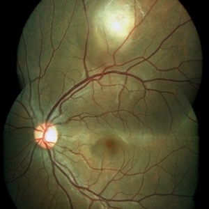
 Fibrotic granuloma vs. Pseudoduplication of the Optic Disc
Nov 29 2023 by Virginia Gebhart
74 year-old female with presumed fibrotic granuloma. Previously diagnosed as pseudoduplication of the optic disc by general ophthalmologist. OCT showed elevation in the RPE, more consistent with granuloma. Pt has been aware for many years, asymptomatic. Will observe.
Photographer: Virginia Gebhart
Imaging device: Topcon 50DX
Condition/keywords: fibrotic scar, granuloma, Pseudoduplication of optic disc
-
 Granulomatous Uveitis
May 18 2020 by McGill University Health Centre
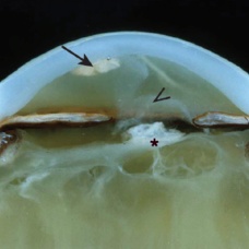
Granulomatous uveitis is found in many inflammatory diseases, and is generally characterized by a predominant histiocytic infiltrate forming a “wall” (granuloma) around a pathogen or foreign body. This is an example of granulomatous uveitis. The eye is aphakic; the uveal track is thickened; and a granuloma is present and attached to the endothelium of the cornea (arrow). The anterior chamber is filled with a hazy material (arrowhead). The vitreous is fibrotic and tractional bands are also present (*).
Condition/keywords: granulomatous uveitis
-
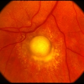
 Ocular Toxocariasis slide 3
Oct 22 2012 by Ronald C. Gentile, MD
The sub-retinal scarred granuloma was white in color and elevated. It had pigment speckling around it. Serum testing was positive for past exposure to Toxocara canis.
Photographer: The New York Eye & Ear Infirmary Department of Medical Imaging
Condition/keywords: toxocariasis
-
 Toxocara Granuloma
Feb 25 2013 by Henry J. Kaplan, MD
Toxocara granuloma superotemporal to the fovea.
Condition/keywords: ocular toxoplasmosis, toxocara granuloma, toxocariasis
-
 Toxocara Granuloma
Feb 25 2013 by Henry J. Kaplan, MD
Toxocara granuloma , epiretinal membrane and localized tractional detachment.
Condition/keywords: toxocara granuloma
-
 Berlin's Nodules
May 2 2013 by Henry J. Kaplan, MD
Granulomatous berlin's nodules in the angle secondary to sarcoidosis.
Condition/keywords: Berlin's nodules, sarcoidosis
-
 Toxocara Granuloma
Feb 25 2013 by Henry J. Kaplan, MD
Toxocara granuloma in the midperiphery of the retina.
Condition/keywords: ocular toxoplasmosis, toxocara granuloma, toxocariasis
-
 Choroidal Granuloma
Apr 23 2019 by Purva Patwari
22-year-old male patient presented with blurring of vision in the right eye noticed since last one week. He was asymptomatic a week ago when he noticed the blurring in his right eye. On examination his vision was 6/6 in both eyes. Anterior segment was normal. Posterior segment was normal for the left eye. Right eye examination revealed a clear vitreous cavity with choroidal granulomas scattered throughout the fundus. The present picture shows choroidal granulomas with OCT segment passing through the parafoveal lesion showing subretinal fluid accumulation and corresponding thickening of the retinal layers. CT scan reveals heterogeneously enhancing lymph nodes showing conglomerationin the hilar region-possibility of tubercular etiology.
Photographer: Dr Purva Patwari, Patwari Retina Center
Imaging device: Zeiss Visu 500
Condition/keywords: choroidal granuloma, choroiditis, granulomatous choroiditis, tubercular choroidal granuloma, tuberculosis
-
 Choroidal Granuloma Secondary to Tuberculosis
Mar 14 2013 by Eduardo Torres-Porras, MD
OCT scan through the granuloma shows attachment of the retinal pigment epithelial-choriocapillaris layer and the neurosensory retina over the granuloma (“contact” sign), inflammatory retinal infiltrate in the deeper retinal layers and subretinal fluid.
Photographer: Eduardo Torres Porras
Imaging device: Cirrus
Condition/keywords: optical coherence tomography (OCT), tubercular choroidal granuloma
-
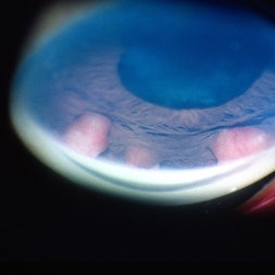
 Busacca nodules
May 2 2013 by Henry J. Kaplan, MD
Typical Busacca iris stromal nodules in sarcoid uveitis; notice the ps formation.
Condition/keywords: busacca nodulaes, granulomatous uveitis, iris nodules, sarcoid bussaca iris nodules
-
 ICG: Choroidal Aspergilloma With Secondary Choroidal Neovascularization and Exudative Retinal Detachment
Mar 21 2019 by Scott D Walter, MD, MSc, FASRS
Multimodal imaging of a transplant patient with disseminated Aspergillosis and vision loss in her left eye.
Condition/keywords: choroidal neovascular membrane (CNVM), choroidal neovascularization (CNV), exudative detachment, focal chorioretinitis, fungal endophthalmitis, granulomatous choroiditis
-
 Bartonella Posterior Granulomatous Mass with Exudative Detachment Post 2 Months
Sep 26 2020 by Swati Agarwal-Sinha, MD, FASRS
Color fundus photo of the left eye two months post-treatment showing resolved exudative detachment, fibrosed granuloma at the optic disc, tractional detachment at the posterior pole and extensive diffuse subretinal exudates extending all around up to the retinal periphery. No retinal vascular changes are noted.
Photographer: Harry Rosa, University of Florida
Condition/keywords: Bartonella bacteria, exudative detachment
-
---thumb.JPG/image-square;max$300,300.ImageHandler) Blastomycosis
Blastomycosis
Oct 28 2012 by Mallika Goyal, MD
Right eye fundus photograph of 32-year-old gentleman shows a large peripapillary choroidal granuloma. Lymph node biopsy had confirmed systemic blastomycosis. The choroidal granuloma resolved with systemic and intravitral voriconazole.
Condition/keywords: blastomycosis, choroidal granuloma
-
 Choroidal Granuloma Secondary to Tuberculosis
Mar 14 2013 by Eduardo Torres-Porras, MD
OCT scan through the granuloma shows attachment of the retinal pigment epithelial-choriocapillaris layer and the neurosensory retina over the granuloma (“contact” sign), inflammatory retinal infiltrate in the deeper retinal layers and subretinal fluid.
Photographer: Eduardo Torres Porras, Laser y ultrasonido ocular de Puebla
Imaging device: Cirrus
Condition/keywords: optical coherence tomography (OCT), tubercular choroidal granuloma
-
 Ocular Toxocariasis slide 1
Oct 22 2012 by Ronald C. Gentile, MD
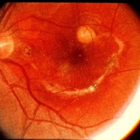
8-year-old boy with a history of puppy exposure failed his school screening in the right eye. Fundus examination revealed a old scarred granuloma involving the macula. Serum testing for anti-Toxocara antibodies were positive.
Photographer: The New York Eye & Ear Infirmary Department of Medical Imaging
Condition/keywords: scarred granuloma, toxocariasis
-
 Peripheral Choroidal Granuloma Associated With Tuberculosis Choroiditis
Jun 3 2017 by S. Natarajan, MD, FASRS, FRCS (GLASGOW) , FICO, D.Sc, FELA
Funds photograoh of an 21-year-old female pheripheral choroidal granuloma associated with tuberculos choroiditis.
Photographer: miss ashwini borde
Imaging device: Carl Zeiss 450 Plus IR
Condition/keywords: peripapillary choroidal granuloma
-
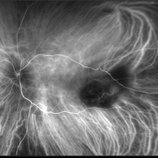
 Toxocara Granuloma
Feb 25 2013 by Henry J. Kaplan, MD
Toxocara granuloma of optic nerve head. Red free image #1; the rest of F/A in the following slides.
Condition/keywords: toxocara granuloma
-
---thumb.JPG/image-square;max$300,300.ImageHandler) Tubercular choroidal granuloma
Tubercular choroidal granuloma
Oct 26 2012 by Mallika Goyal, MD
Fundus photograph of left eye of a 43-year-old HIV infected gentleman who started antiretroviral therapy 2 months prior to visual symptoms in this eye.
Condition/keywords: tubercular choroidal granuloma
 Loading…