Search results (453 results)
-
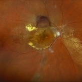
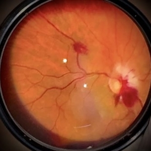
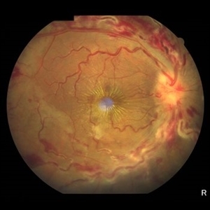
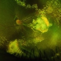
 Peripheral Exudative Hemorrhagic Chorioretinopathy
Dec 17 2025 by Virginia Gebhart
77 year old female with amelanotic choroidal lesion with subretinal hemorrhage at the base and exudate inferiorly. Lesion has decreased on exam, photos and ultrasound after 4 months. Ultrasound showed irregular lesion that is regressing, variable internal reflectivity, and no venous pulsations. Findings most consistent with PEHCR. Will continue to observe.
Photographer: Virginia Gebhart, Retina Consultants of Carolina
Imaging device: Optos California
Condition/keywords: asteroid hyalosis, peripheral exudative hemorrhagic chorioretinopathy (PEHCR)
-
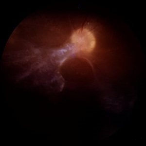
 Racemose Hemangioma
Dec 16 2025 by Seif Allah Anwar
18 year-old female with dilated, tortuous arteriovenous communication without an intervening capillary bed. Vessels may appear coiled or spaghetti-like extending into the foveal region with no associated retinal hemorrhages, exudates, or edema On OCT : the anomalous vessels appear hyper reflective spanning the whole retinal thickness with ILM draping, No associated subretinal or intraretinal fluid.
Photographer: Seif Anwar , KING SALMAN INTERNATIONAL UNIVERSITY
Imaging device: TOPCON
Condition/keywords: racemose hemangioma
-
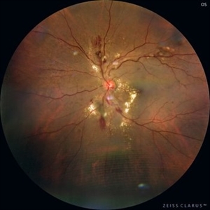
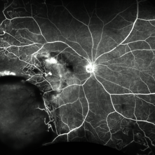
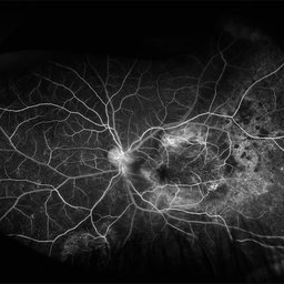
 Leber’s Miliary Aneurysm
Dec 12 2025 by KANWALJEET HARJOT MADAN, M.S. (Ophthalmology); FAICO (Vitreous - Retina)
A 34 year-old male presented with decrease vision in right eye for 3 months. Anterior segment exam was normal. Fundus exam in RE revealed presence of macular edema which was evident on OCT. Multiple retinal vascular aneurysmal dilatations with telangiectasia of the retina blood vessels noted superiorly which was evident on FFA. These aneurysms were multiple, tiny and leaky on FFA. He was diagnosed to have Leber’s miliary aneurysms. It is a rare, typically unilateral eye condition, often seen in young males, characterized by multiple tiny, leaky aneurysms in the retinal blood vessels, leading to deposits of hard exudates and potential vision loss, especially if it affects the macula. It is considered a milder form of Coats' disease.
Photographer: Dr. Kanwaljeet Harjot Madan, Thind Eye Hospital, Jalandhar City (Punjab) INDIA.
Imaging device: Zeiss Fundus Camera
Condition/keywords: FFA, Leber's miliary aneurysm
-
 Cracking the Angioid Streaks Mystery
Nov 26 2025 by SHRADDHA RAJ SHRIVASTAVA
Left eye pseudocolor fundus photo showing hyperpigmented irregular lines emanating from the disc in a radiating fashion. Surrounding the angioid streaks and at the posterior pole, we can see numerous dot-like hypopigmented deposits along with a grayish-green membrane with exudates at the macula. The image is suggestive of Angioid Streaks with CNVM.
Photographer: Dr. Shraddha Raj Shrivastava
Imaging device: Nidek Mirante SLO/OCT (Confocal scanning/Spectral domain OCT)
Condition/keywords: Angiod streaks in Pseudoxanthoma elasticum, Angioid Streaks, Bruch's membrane, choroidal neovascular membrane (CNVM), color fundus photograph, pseudoxanthoma elasticum (PXE)
-
 Neovascular Medusa: A Bad Hair Day at the Optic Disc
Nov 4 2025 by SHRADDHA RAJ SHRIVASTAVA
Left eye pseudocolor fundus photo of 67 year old male, diagnosed with both eyes proliferative diabetic retinopathy, showing hair-like fronds of active neovascularisation at the disc (NVD) extending into the vitreous, giving the medusa-head appearance. There is a band of fibrovascular proliferation nasal to the disc, with presence of hard exudates and dot hemorrhages at the macula.
Photographer: Dr. Shraddha Raj Shrivastava
Imaging device: Nidek Mirante SLO/OCT (Confocal scanning/Spectral domain OCT
Condition/keywords: Diabetic Retinopathy, fibrovascular proliferation, Neovascularisation at the Disc (NVD), proliferative diabetic retinopathy (PDR)
-
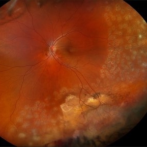
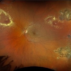
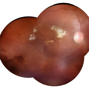
 All That Glows Yellow Isn’t Mellow: Coats' Disease Unveiled
Nov 4 2025 by SHRADDHA RAJ SHRIVASTAVA
Montage fundus image of an 11 year old boy diagnosed with left eye Coats' disease (stage 3A1), reveals a hyperemic disc and surrounding intra-retinal exudates superior to the disc. There is a single fibroglial nodule at the macula causing submacular fibrosis with exudation. We can see areas of pigmentary changes and RPE atrophy in posterior pole and mid-peripheral retina supero-temporally. There is massive yellowish subretinal exudation in all the quadrants, which are associated with telangiectatic aneurysmal capillary dilation, more prominently seen in the nasal periphery. Supero-nasally we can also see an orange-red elevated vaso-proliferative mass with overlying dilated capillaries, which has likely developed secondary to untreated long standing disease. We can also see associated extrafoveal subtotal exudative retinal detachment in the inferior and nasal quadrants.
Photographer: Dr. Shraddha Raj Shrivastava
Imaging device: Nidek Mirante SLO/OCT (Confocal scanning/Spectral domain OCT)
Condition/keywords: COATS DISEASE, exudative detachment, leukocoria, subretinal exudates, Xanthocoria, yellow exudate
-
 Proliferative Ring of Fire
Oct 29 2025 by SHRADDHA RAJ SHRIVASTAVA
Right eye color fundus photo of 57 year old male, diagnosed with both eyes high risk proliferative diabetic retinopathy (PDR). Posterior pole reveals Neovascularization of disc (NVD) with extensive fibrovascular proliferations (FVPs) overlying the disc and along the arcades. We can also see a florid network of neovascularization (NVEs), with veins showing looping and beading changes. Hard exudates and dot-blot hemorrhages were seen at the macula.
Photographer: Dr. Shraddha Raj Shrivastava
Imaging device: Nidek Mirante SLO/OCT (Confocal scanning/Spectral domain OCT)
Condition/keywords: fibrovascular proliferation, Neovascularisation elsewhere (NVE), NVE, proliferative diabetic retinopathy (PDR), venous beading
-
 Neovascular AMD
Oct 3 2025 by Kimberly Wakester
Optomap RGB of an 80-year-old-woman with Neovascular AMD in the right eye. There is recurrent migration of fluid and exudate inferiorly on exam. There is also a superior hemorrhagic PED. Last Avastin treatment was in 2015. Recommended treatment in her right eye with Avastin. Patient will return for follow up care and repeat OCT.
Photographer: Kimberly Wakester, COA, OCT-C
Imaging device: Optos California
Condition/keywords: Hemorrhagic PED, migration of fluid, Neovascular AMD
-
 Hypertensive Retinopathy
Sep 16 2025 by Píndaro Alonso Cruz-Benitez
Hypertensive Retinopathy Stage 3 Male 55 year-old
Photographer: Pindaro Alonso Cruz Benitez
Condition/keywords: exudate, Haemorrhage, hypertensive retinopathy
-
 Retinal Artery Macroaneurysm With Macular Edema
Sep 12 2025 by Tejaswita Verma
Fundus photo of a 73 year old hypertensive female with 6/18 vision, presenting with RAM ,with surrounding hard exudates and macular edema. She was advised focal laser, anti VEGF injection.
Photographer: DR. TEJASWITA VERMA
Imaging device: MIRANTE
Condition/keywords: RAM, retinal arterial macroaneurysm
-
 Macular Tributary Retinal Venous Occlusion
Sep 7 2025 by Anand Temkar
A 55 yrs old female, k/c/o DM ( type II ) since past 5 yrs ( on medication ). Her vision was 6/6 in RE and 6/24 in her LE. IOP was 16 in both eyes. On examination, RE was WNL, and in LE ( color photo ) we noticed exudates, small hemorrhages, edema and sclerosed vessel ( depicted by black arrow. OCT LE shows altered foveal contour with cystic spaces and intraretinal hyperreflective material ( IRHRM ).
Photographer: Dr.Anand Temkar- Vasan Eye Hospital, Tiruchirappalli
Imaging device: Opticon
Condition/keywords: macular branch retinal vein occlusion (BRVO), venous occlusion
-
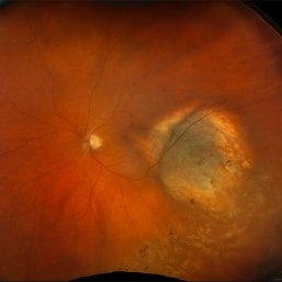
 Vasoproliferative Tumor (FEVR) s/p PPV/PRP
Aug 27 2025 by Virginia Gebhart
39 year old female with an amelanotic vascular lesion inferotemporal with CR atrophy inferior edge and likely lipid exudate superior edge. Pt presented with vitreous and sub-hyaloid hemorrhage. Findings from exam, ultrasound, FA all consistent with FEVR, stage 2. PPV with PRP performed, pt vison has improved from CF@2ft at initial visit to 20/100 PH 20/60 at 1 week post-op. Pt's 2 children have been recently examined with identical findings of FEVR
Photographer: Virginia Gebhart, Retina Consultants of Carolina
Imaging device: Optos California
Condition/keywords: familial exudative vitreoretinopathy (FEVR), pan-retinal photocoagulation (PRP), Vasoproliferative Tumor
-
 Retinal Macroaneurysm OS (RAM)
Aug 20 2025 by Drew Mitchell
Optos Color photograph of a 79 year old woman with non central macular edema and exudates around RAM inferotemporally.
Photographer: Drew Mitchell OCT-C
Imaging device: Optos Silverstone
Condition/keywords: color photo, exudates, OPTOS, RAM, retinal macroaneurysm
-
 Bilateral Roth Spot in the Setting of Mitral Valve Endocarditis
Aug 18 2025 by Helder Vasconcelos
A 55-year-old man with chronic alcoholism presented with wasting and fever. The symptoms were preceded by a recent tooth extraction and gingivitis. Fundus examination in the ICU showed a retinal hemorrhage with a white spot (Roth spot) associated with peripapillary hemorrhage and cotton wool exudate. A similar Roth spot was observed in the contralateral eye.
Photographer: Helder Vasconcelos
Imaging device: Smartphone Fundoscopy
Condition/keywords: Infectious endocarditis, Roth Spots
-
 Retinal Aneurysms
Aug 6 2025 by Korey Starkey
54 year-old patient presents with scattered peripheral aneurysms with exudates. FA was performed showing peripheral nonperfusion and aneurysms. Treated patient with PRP and focal laser to aneurysms and continued observation.
Photographer: Kore Starkey
Imaging device: Optos
Condition/keywords: aneurysm, branch retinal vein occlusion (BRVO), chorioretinal scar, circinate ring, exudates, fundus photography, lesion, Optos, retinal aneurysms
-
 New Choroidal Melanoma
Jul 16 2025 by Virginia Gebhart
78 year old male with a partially amelanotic dome-shaped lesion with RPE changes, hard exudates, overlying intraretinal fluid and minimal SRF temporally. Exam and ultrasound findings consistent with choroidal melanoma. Pt will be scheduled for brachytherapy pending CT scan results.
Photographer: Virginia Gebhart, Retina Consultants of Carolina
Imaging device: Optos California
Condition/keywords: amelanotic melanoma, choroidal melanoma
-
 Neuroretinitis
Jul 7 2025 by César Adrián Gómez Valdivia, MD
Neuroretinitis found in a 38 YO male patient with IV drugs abuse history. Findings were bilateral. The lipid-rich component of the exudate is able to penetrate into the outer plexiform layer, creating what is clinically seen as a macular star pattern.
Photographer: @eyemissu2
Imaging device: TOPCON TRC-50DX
Condition/keywords: neuroretinitis
-
 Fluorescein Angiography (FA) of a Primary Retinal Vasoproliferative Tumor
Jun 29 2025 by Marcelo Zas, MD PhD
We present a case of a 33-year-old male patient, who presented with decreased visual acuity in his right eye with 20/80, presenting a primary retinal vasoproliferative tumor in the lower temporal quadrant. The fluorescein angiography findings are: 1. Early hyperfluorescence due to its rich intrinsic vascularity and often has dilated feeding arterioles and draining venules. 2. Marked progressive leakage from the tumor vessels. 3. The late leakage often obscures fine vascular details in the late phase and corresponds to exudation and macular edema seen clinically. 4. Staining of surrounding exudates, RPE disturbances and gliosis. 5. In our case also a marked peripheral capillary closure in the areas adjacent to the tumor and in other quadrants as well.
Photographer: Marcelo Zas MD PhD
Condition/keywords: RETINAL VASOPROLIFERATIVE TUMOR
-
 Retinal Vasoproliferative Tumor
Jun 24 2025 by Marcelo Zas, MD PhD
We present a case of a 33-year-old male patient, who presented with decreased visual acuity in his right eye with 20/80, presenting a primary retinal vasoproliferative tumor in the lower temporal quadrant. The tumor is associated with serous retinal detachment, hard exudation, neovascularization and telangiectasias. Lipid exudates extend toward the macula, indicating macular involvement, which may contribute to decreased visual acuity. Oi was normal with 20/20 of BCVA. The patient was treated initially with IV anti-VEGF therapy and cryotherapy.
Photographer: Marcelo Zas MD PhD
Condition/keywords: RETINAL VASOPROLIFERATIVE TUMOR
-
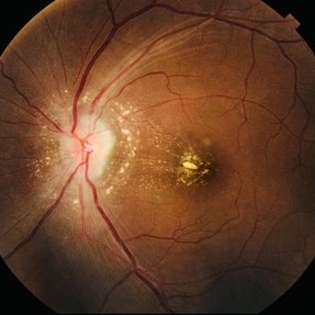
 Central Retinal Vein Occlusion
Jun 21 2025 by Moazzam Parvez
Fundus photograph of a 56 year old male presenting with dilated tortuous vessels with adjoining Hard exudates and macular star.
Photographer: Moazzam Parvez , Netralayam , Kolkata
Imaging device: Topcon Maestro 2
Condition/keywords: CRVO with macular edema, hard exudates, macular star
-
 Diabetic Retinopathy
Jun 4 2025 by Paulina Araujo
The 55-degree central fundus photograph of the right eye demonstrates numerous hard exudates, dot intraretinal hemorrhages, and microaneurysms.
Photographer: Paulina D.Araujo Martínez, Asociación para Evitar la Ceguera en México I.A.P., Hospital Dr Luis Sánchez Bulnes.
Condition/keywords: diabetic retinopathy
-
 Macular Edema
Jun 4 2025 by Paulina Araujo
The composite fundus photograph of the right eye demonstrates circinate hard exudates in the thickened macular area, along with flame-shaped intraretinal hemorrhages along the inferior temporal arcade.
Photographer: Paulina D.Araujo Martínez, Asociación para Evitar la Ceguera en México I.A.P., Hospital Dr Luis Sánchez Bulnes.
Condition/keywords: macular edema
-
 Tractional Retinal Detachment
Jun 4 2025 by Paulina Araujo
The 55-degree central fundus photograph of the right eye reveals a thickened and opacified hyaloid exerting traction on the optic disc and posterior pole of the retina, along with hard exudates and microaneurysms consistent with advanced proliferative diabetic retinopathy.
Photographer: Paulina D.Araujo Martínez, Asociación para Evitar la Ceguera en México I.A.P., Hospital Dr Luis Sánchez Bulnes.
Condition/keywords: tractional retinal detachment
-
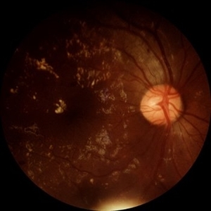
 Coats Disease
May 27 2025 by César Adrián Gómez Valdivia, MD
Fluorescein Angiography on an 8 year-old male patient with Coats disease. Vascular leakage causes hard exudates which may be peripheral (near the vascular abnormalities) or midperipheral and central (at the macula. Findings were bilateral.
Photographer: @eyemissu2
Imaging device: California ICG OPTOS
Condition/keywords: Coats disease
-
 Coats Disease
May 27 2025 by César Adrián Gómez Valdivia, MD
Fundus photograph of an 8 year-old male patient with Coats disease. Vascular leakage causes hard exudates which may be peripheral (near the vascular abnormalities) or midperipheral and central (at the macula). Findings were bilateral.
Photographer: @eyemissu2
Imaging device: California ICG OPTOS
Condition/keywords: Coats disease
 Loading…