Search results (453 results)
-
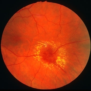
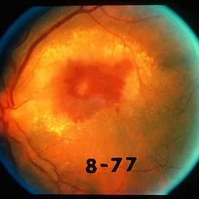
 Cytomegalovirus Retinitis with Severe Hemorrhage and Exudate Right Eye
Oct 9 2012 by Jeffrey G. Gross, MD, FASRS
CMV retinitis with severe hemorrhage and exudate, right eye.
Condition/keywords: exudate
-
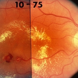
 Branch Retinal Vein Occlusion with Macular Edema
Aug 23 2012 by Gerardo Garcia-Aguirre, MD
Fundus photograph composition of the left eye, showing flame-shaped and blot hemorrhages in the superotemporal quadrant, with hard exudates surrounding the fovea.
Photographer: Noemí Hernández, Asociación para Evitar la Ceguera en México
Condition/keywords: branch retinal vein occlusion (BRVO), macular edema
-
 Hypertensive Retinopathy
Feb 25 2013 by Suber S. Huang, MD, MBA, FASRS
32-year-old African American male with Grade IV hypertensive retinopathy and acute renal failure. Vision OD 20/70, OS 20/25. Creatine 7.1. BP: 250/150.
Photographer: Geoffrey Pankhurst, University Hospitals, Eye Institute/Dept. Ophthalmology and Visual Sciences Case Western Reserve University Cleveland, OH
Imaging device: Topcon TRC 50x
Condition/keywords: acute renal failure, disc edema, exudate, hypertension, hypertensive retinopathy, ischemia, macular edema, macular ischemia, optic disc edema
-
 Retinal arterial macroaneurysm
Jan 11 2013 by Alex P. Hunyor, MD
Retinal arterial macroaneurysm with associated haemorrhage and surrounding lipid exudate.
Condition/keywords: macroaneurysm, retinal arterial macroaneurysm
-
 Arterial Macroaneurysm
Mar 29 2013 by Henry J. Kaplan, MD
Typical arterial macroaneurysm surrounded by lipid exudates and edema.
Condition/keywords: macroaneurysm, retinal arterial macroaneurysm
-
 Hypertensive Retinopathy
Feb 25 2013 by Suber S. Huang, MD, MBA, FASRS
32-year-old African American male with Grade IV hypertensive retinopathy and acute renal failure. Vision OD 20/70, OS 20/25. Creatine 7.1. BP: 250/150.
Photographer: Geoffrey Pankhurst, University Hospitals, Eye Institute/Dept. Ophthalmology and Visual Sciences Case Western Reserve University Cleveland, OH
Imaging device: Topcon TRC 50x
Condition/keywords: acute renal failure, disc edema, exudate, hypertension, hypertensive retinopathy, ischemia, macular edema, macular ischemia, optic disc edema
-
---thumb.JPG/image-square;max$300,300.ImageHandler) hypertensive retinopathy
hypertensive retinopathy
Nov 3 2012 by Mallika Goyal, MD
Left eye of a 30-year-old hypertensive gentleman shows hypertensive retinopathy grade 4 with retinal hemorrhages, exudates and disc edema.
Photographer: Mallika Goyal, MD
Condition/keywords: disc edema, hypertensive retinopathy, retinal hemorrhage
-
 Diabetic Retinopathy Hard Exudates OS
Jun 30 2013 by Rogerio N Shinsato, MD, PhD
Fundus photograph with diabetic retinopathy.
Condition/keywords: diabetic macular edema, foveal hard exudates
-
 Diabetic Retinopathy, CSME, Color Fundus Photo
Mar 18 2015 by James B. Soque, CRA, OCT-C, COA, FOPS
A 58-year-old diabetic male with a longstanding history of diabetic eye disease. Left eye color fundus photo shows extensive CSME, Clinically Significant Macular Edema, with deposits of hard exudates at fixation. There is extensive scattering of hard exudates and sheathing of the vessels.
Photographer: James B Soque, CRA COA
Imaging device: Topcon TRC 50 DX, OIS 5 MP Camera, MERGE software
Condition/keywords: background diabetic retinopathy (BDR), creamy yellow exudates, diabetes, exudates over the posterior pole, neovascularization of the disc (NVD), vessel sheathing
-
---thumb.JPG/image-square;max$300,300.ImageHandler) hypertensive retinopathy
hypertensive retinopathy
Nov 3 2012 by Mallika Goyal, MD
Right eye of a 30-year-old hypertensive gentleman shows hypertensive retinopathy grade 4 with retinal hemorrhages, exudates and disc edema.
Photographer: Mallika Goyal, MD
Condition/keywords: disc edema, hypertensive retinopathy, retinal hemorrhage
-
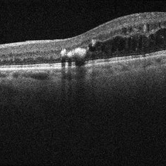 Circinate Ring OCT
Aug 24 2012 by John S. King, MD
DR; hard exudate at edges of macular edema and are hyper-reflective with shadowing.
Photographer: Kristin Konecki, OcuSight Eye Care Center, Rochester, NY
Condition/keywords: circinate ring
-
 Pars Planitis - Peripheral Uveitis
Nov 9 2012 by Norman Byer
This 25-year-old man had pars planitis, peripheral uveitis bilaterally. In this eye it produced a small tractional oval tear of the retina and an inferior retinal detachment. The typical creamy yellow exudates of pars planitis can be seen in the lower right very close to the ora serrata.
Condition/keywords: creamy yellow exudates, inferior retinal detachment, pars planitis, peripheral uveitis, tractional retinal tear
-
 Leukemia
Feb 13 2013 by From the Collections of Thomas M. Aaberg, MD and Thomas M. Aaberg Jr., MD
White centered hemorrhage, ischemia exudate.
Condition/keywords: exudate, ischemia, leukemia
-
---thumb.jpg/image-square;max$300,300.ImageHandler) Diabetic Retinopathy Hard Exudates OD
Diabetic Retinopathy Hard Exudates OD
Jun 30 2013 by Rogerio N Shinsato, MD, PhD
Fundus photograph with diabetic retinopathy.
Condition/keywords: diabetic macular edema, foveal hard exudates
-
 Papilledema
Sep 21 2012 by Suber S. Huang, MD, MBA, FASRS
Fundus photograph of a 24-year-old obese woman with severe papilledema secondary to idiopathic intracranial hypertension.
Condition/keywords: dilated tortuous vessels, exudate, idiopathic intracranial hypertension, Paton's lines, peripapillary hemorrhage, pseudotumor cerebri
-
 Branch Retinal Vein Occlusion with Diffuse Lipid Exudate
Oct 16 2012 by Jeffrey G. Gross, MD, FASRS
BRVO with diffuse lipid exudate.
Condition/keywords: branch retinal vein occlusion (BRVO), diffuse lipid exudate
-
 Leukemia
Feb 13 2013 by From the Collections of Thomas M. Aaberg, MD and Thomas M. Aaberg Jr., MD
White centered hemorrhage, ischemia exudate.
Condition/keywords: exudate, ischemia, leukemia
-
 Arterial Macro Aneurysm
Mar 29 2013 by Henry J. Kaplan, MD
Large arterial macroaneurysm with hemorrhage and surrounding exudates.
Condition/keywords: retinal arterial macroaneurysm
-
 Diabetic Retinopathy, CSME, Exudates, NVD, Color Fundus Photo, Montage
Mar 18 2015 by James B. Soque, CRA, OCT-C, COA, FOPS
A 58-year-old diabetic male with a longstanding history of diabetic eye disease. Left eye color fundus photo shows extensive CSME, Clinically Significant Macular Edema, with deposits of hard exudates at fixation. There is extensive scattering of hard exudates and sheathing of the vessels.
Photographer: James B Soque, CRA COA
Imaging device: Topcon TRC 50 DX, OIS 5 MP Camera, MERGE software
Condition/keywords: background diabetic retinopathy (BDR), creamy yellow exudates, diabetes, exudates over the posterior pole, neovascularization of the disc (NVD), vessel sheathing
-
---thumb.jpg/image-square;max$300,300.ImageHandler) Yellow White Subretinal Exudates
Yellow White Subretinal Exudates
Feb 13 2013 by From the Collections of Thomas M. Aaberg, MD and Thomas M. Aaberg Jr., MD
Yellow white subretinal exudates/lesions in macula and mid periphery.
Condition/keywords: macula, subretinal exudates
-
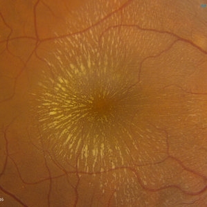 Cat Scratch
Feb 15 2017 by Hua Gao, MD, PhD, FASRS
A female patient of 57-year-old presented with neuroretinitis due to cat-scratch disease with positive Bartonella henselae antibodies. Two weeks after symptom onset she developed exudates in a "macular star" pattern.
Condition/keywords: cat scratch retinitis
-
 Type 1A Macular Telangiectasia - Fundus photograph
Nov 11 2013 by Gerardo Garcia-Aguirre, MD
Fundus photograph of a 43-year-old male complaining of mild metamorphopsia in OS. BCVA 20/25. Some hard exudates and telangiectatic vessels are observed inferior and temporal to the fovea.
Condition/keywords: macular telangiectasia
-
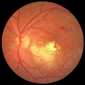
---thumb.JPG/image-square;max$300,300.ImageHandler) Diabetic Macular Edema
Diabetic Macular Edema
Oct 26 2012 by Mallika Goyal, MD
Fundus photograph of left eye of 55-year-old diabetic and hypertensive gentleman with normal serum lipids showing abundant foveal hard exudates.
Condition/keywords: diabetic macular edema
-
 Hypertensive Retinopathy
Dec 12 2012 by Mallika Goyal, MD
Left eye of a 35-year-old hypertensive gentleman with BP 240/120 mm Hg when first detected a week prior to presentation. Retinal haemorrhages, exudates, macular star and disc edema suggestive of hypertensive retinopathy grade 4.
Photographer: Mallika Goyal, MD, Apollo Hospitals, Hyderabad, India
Condition/keywords: hypertensive retinopathy
-
---thumb.JPG/image-square;max$300,300.ImageHandler) diabetic macular edema
diabetic macular edema
Oct 26 2012 by Mallika Goyal, MD
Fundus photograph of left eye of 58-year-old diabetic gentleman with normal serum lipids showing foveal hard exudates.
Condition/keywords: foveal hard exudates
 Loading…