Search results (453 results)
-
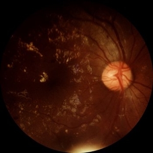
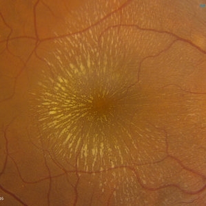 Cat Scratch
Feb 15 2017 by Hua Gao, MD, PhD, FASRS
A female patient of 57-year-old presented with neuroretinitis due to cat-scratch disease with positive Bartonella henselae antibodies. Two weeks after symptom onset she developed exudates in a "macular star" pattern.
Condition/keywords: cat scratch retinitis
-
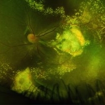
 Coats' Disease
Feb 2 2021 by Niloofar Piri, MD
#2 Fluorescein angiography of the same patient in lamellar arteriovenous phase, demonstrating temporal peripheral telangiectatic vessels, as well as hyperfluorescent aneurysma lesions. Note the anterior capillary non perfusion. Posterior hypofluorescence is secondary to blocking effect from hard exudates.
Condition/keywords: Coats' disease, Leber's miliary aneurysm
-
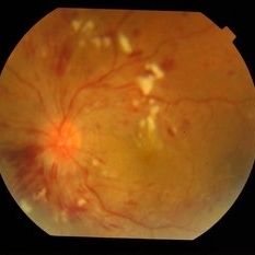
 Hypertensive Retinopathy
Feb 25 2013 by Suber S. Huang, MD, MBA, FASRS
32-year-old African American male with Grade IV hypertensive retinopathy and acute renal failure. Vision OD 20/70, OS 20/25. Creatine 7.1. BP: 250/150.
Photographer: Geoffrey Pankhurst, University Hospitals, Eye Institute/Dept. Ophthalmology and Visual Sciences Case Western Reserve University Cleveland, OH
Imaging device: Topcon TRC 50x
Condition/keywords: acute renal failure, disc edema, exudate, hypertension, hypertensive retinopathy, ischemia, macular edema, macular ischemia, optic disc edema
-
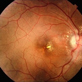
 Arterial Macroaneurysm
Mar 29 2013 by Henry J. Kaplan, MD
Typical arterial macroaneurysm surrounded by lipid exudates and edema.
Condition/keywords: macroaneurysm, retinal arterial macroaneurysm
-
 Branch Retinal Vein Occlusion with Macular Edema
Aug 23 2012 by Gerardo Garcia-Aguirre, MD
Fundus photograph composition of the left eye, showing flame-shaped and blot hemorrhages in the superotemporal quadrant, with hard exudates surrounding the fovea.
Photographer: Noemí Hernández, Asociación para Evitar la Ceguera en México
Condition/keywords: branch retinal vein occlusion (BRVO), macular edema
-
 Central Retinal Vein Occlusion
Jun 21 2025 by Moazzam Parvez
Fundus photograph of a 56 year old male presenting with dilated tortuous vessels with adjoining Hard exudates and macular star.
Photographer: Moazzam Parvez , Netralayam , Kolkata
Imaging device: Topcon Maestro 2
Condition/keywords: CRVO with macular edema, hard exudates, macular star
-
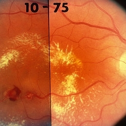
 Coats' Disease
Feb 25 2021 by Niloofar Piri, MD
Collage color photo and FA image of the same patient with Coats' Disease demonstrating telangiectatic aneurysmal lesions in the temporal periphery, associated with hard exudate deposition posteriorly. FA (AV phase) demonstrating hyperfluorescent aneurysmal lesions as well as peripheral capillary non perfusion. Note the posterior hypofluorescence where the hard exudates are located.
Condition/keywords: Coats' disease, congenital retinal telangiectasis, retinal telangiectasia
-
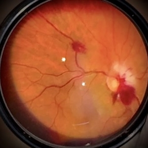
 Coats' Disease
Feb 2 2021 by Niloofar Piri, MD
#1 16-year-old male with abnormal temporal peripheral telangiectatic and aneurysmal vascular lesions associated with hard exudate deposition posteriorly. Vision 20/20. Stage II Coats' disease.
Condition/keywords: Coats' disease, Leber's miliary aneurysm
-
 Coats' Disease - Stage 3A
Aug 21 2019 by Victor M Villegas, MD
Coats' Disease - stage 3A.
Condition/keywords: abnormal retina, Coats' disease, diffuse lipid exudate, edema, foveal hard exudates, pediatic retina, retcam, retinal angioma
-
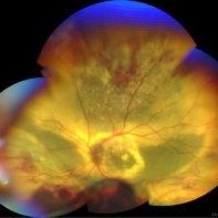
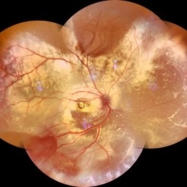 Coats' Disease Montage
Feb 5 2021 by Akansha Sharma
Fundus photograph of a 5-year-old male child who presented with unilateral diminution of vision since one month.
Photographer: Dr. Nivesh Gupta, M.S., Retina Foundation, Ahmedabad
Condition/keywords: angiomatosis retinae, Coats' disease, exudative detachment, subretinal exudates
-
 Acute Necrotizing Retinal Vasculitis as Onset of Systemic Lupus Erythematosus.
Sep 3 2016 by ADRIANO FERREIRA
A 28-year-old white man was referred to the rheumatology clinic with gradually and rapid deterioration of the vision (both eyes). In this picture, we can observe cotton wool spots in the papillomacular area and extensive hemorrhages in posterior polo and in the middle periphery. Hard exudates are present in macular area (macular edema)
Photographer: Claudio Zett Lobo
Imaging device: TRC50DXi TOPCON
Condition/keywords: systemic lupus erythematosus (SLE) vasculitis, vasculitis
-
 Bilateral Roth Spot in the Setting of Mitral Valve Endocarditis
Aug 18 2025 by Helder Vasconcelos
A 55-year-old man with chronic alcoholism presented with wasting and fever. The symptoms were preceded by a recent tooth extraction and gingivitis. Fundus examination in the ICU showed a retinal hemorrhage with a white spot (Roth spot) associated with peripapillary hemorrhage and cotton wool exudate. A similar Roth spot was observed in the contralateral eye.
Photographer: Helder Vasconcelos
Imaging device: Smartphone Fundoscopy
Condition/keywords: Infectious endocarditis, Roth Spots
-
 Choroidal Melanoma With Radiation Retinopathy
Jul 8 2013 by Jason S. Calhoun
Patient came with follow up on choroidal melanoma. Right eye that was treated back in June of 2009 with a radioactive implant. Vein occlusion is also present with VA - hand motion. Hemorrhages visible with hard exudates from the radiation retinopathy.
Photographer: Jason S. Calhoun, Department of Ophthalmology, Mayo Clinic Jacksonville, Florida
Condition/keywords: radiation retinopathy
-
 Coats Disease
May 27 2025 by César Adrián Gómez Valdivia, MD
Fundus photograph of an 8 year-old male patient with Coats disease. Vascular leakage causes hard exudates which may be peripheral (near the vascular abnormalities) or midperipheral and central (at the macula). Findings were bilateral.
Photographer: @eyemissu2
Imaging device: California ICG OPTOS
Condition/keywords: Coats disease
-
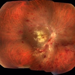
 Coats' disease
Sep 7 2022 by Niloofar Piri, MD
Total exudative RD with extensive subretinal exudates and peripheral telangiectatic vascular anomalies in stage 4 Coats's disease. Patient is a 12 yo who presented with severe eye pain and neovascular glaucoma secondary to the above.
Photographer: Jacob Grodsky, MD
Condition/keywords: Coats' disease
-
 Diabetic Retinopathy
Jun 4 2025 by Paulina Araujo
The 55-degree central fundus photograph of the right eye demonstrates numerous hard exudates, dot intraretinal hemorrhages, and microaneurysms.
Photographer: Paulina D.Araujo Martínez, Asociación para Evitar la Ceguera en México I.A.P., Hospital Dr Luis Sánchez Bulnes.
Condition/keywords: diabetic retinopathy
-
---thumb.jpg/image-square;max$300,300.ImageHandler) Diabetic Retinopathy Hard Exudates OD
Diabetic Retinopathy Hard Exudates OD
Jun 30 2013 by Rogerio N Shinsato, MD, PhD
Fundus photograph with diabetic retinopathy.
Condition/keywords: diabetic macular edema, foveal hard exudates
-
 Diabetic Retinopathy Hard Exudates OS
Jun 30 2013 by Rogerio N Shinsato, MD, PhD
Fundus photograph with diabetic retinopathy.
Condition/keywords: diabetic macular edema, foveal hard exudates
-
 Diabetic Retinopathy Hard Exudates OS
Jun 30 2013 by Rogerio N Shinsato, MD, PhD
Fundus photograph with diabetic retinopathy.
Condition/keywords: diabetic macular edema, foveal hard exudates
-
 Diabetic Retinopathy, CSME, Exudates, NVD, Color Fundus Photo, Montage
Mar 18 2015 by James B. Soque, CRA, OCT-C, COA, FOPS
A 58-year-old diabetic male with a longstanding history of diabetic eye disease. Left eye color fundus photo shows extensive CSME, Clinically Significant Macular Edema, with deposits of hard exudates at fixation. There is extensive scattering of hard exudates and sheathing of the vessels.
Photographer: James B Soque, CRA COA
Imaging device: Topcon TRC 50 DX, OIS 5 MP Camera, MERGE software
Condition/keywords: background diabetic retinopathy (BDR), creamy yellow exudates, diabetes, exudates over the posterior pole, neovascularization of the disc (NVD), vessel sheathing
-
 Eales Disease
May 23 2021 by Katia Delalibera Pacheco, MD
Color fundus photograph of the left eye of a 37-year-old man with Eales disease. Note the peripheral to mid-peripheral periphlebitis in multiple quadrants concurrently. Venous dilation and perivascular exudate can be observed. We can also note the demarcation between perfused and nonperfused retina.
Photographer: CBV- Eye Hospital Brasilia, DF, Brazil
Condition/keywords: Eales disease
-
 Hypertensive Retinopathy With Bilateral Serous Retinal Detachment at Macula
Jul 29 2014 by Mallika Goyal, MD
Left eye fundus of a 36-year-male with sudden vision drop shows grade 4 hypertensive retinopathy with retinal hemorrhages, exudates and ischaemic disc edema. OCT revealed serous retinal detachment at macula.
Photographer: Mallika Goyal, MD, Apollo Health City, Jubilee Hills, Hyderabad-500033
Condition/keywords: hypertensive retinopathy
-
 Idiopathic retinal vasculitis, aneurysms and neuroretinitis
Apr 24 2022 by Aniruddha K Agarwal, MD
Ultra-wide field fundus fluorescein angiography (FFA) of the left eye from an asymptomatic, healthy 33-year-old woman who was referred to the retina clinic from a refractive surgery unit due to the presence of vascular anomalies and hard exudates in both eyes. FFA revealed the characteristic sacular aneurysms at the bifurcation of retinal arterioles in the posterior pole, together with microvascular anomalies and capillary closure peripherally.
Photographer: Julio J GONZALEZ-LOPEZ, MD, PhD, FEBO and Teresa GONZALEZ-LOMAS, RN
Imaging device: Optos California
Condition/keywords: IRVAN Syndrome, IUSG, neuroretinitis, retinal vasculitis, uveitis
-
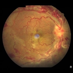
 Infectious Neuroretinitis
May 26 2025 by César Adrián Gómez Valdivia, MD
Neuroretinitis found in a 38 year-oldmale patient with IV drugs abuse history. Findings were bilateral. The lipid-rich component of the exudate is able to penetrate into the outer plexiform layer, creating what is clinically seen as a macular star pattern.
Photographer: @eyemissu2
Imaging device: TOPCON TRC-50DX
Condition/keywords: neuroretinitis
-
 Leber's Miliary Aneurysm
Jan 23 2025 by Tejaswita Verma
A 41 year old male presented with 6/9 vision in the RE and fundus picture revealed miliary aneurysm with exudates and hemorrhages surrounded by old focal and sectoral laser marks. OCT revealed altered foveal contour with cystic spaces and IRHRM. He was advised RE injection antiVEGF with focal laser.
Photographer: DR. TEJASWITA VERMA
Imaging device: MIRANTE
Condition/keywords: Leber's miliary aneurysm
 Loading…