Search results (117 results)
-
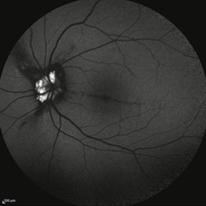
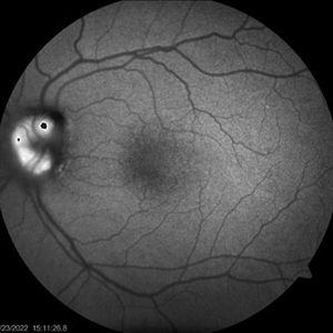
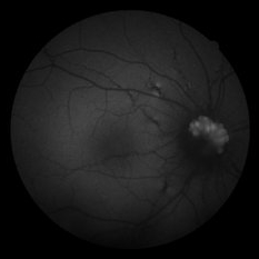
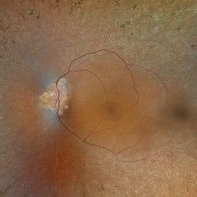 Optic Disc Drusen, RP
Apr 21 2025 by Virginia Gebhart
28 year old male with stable retinitis pigmentosa and optic disc drusen OU. Bardet-Biedl variant identified in previous genetic testing. BCVA 20/50 OD, 20/30 OS
Photographer: Virginia Gebhart, Retina Consultants of Carolina
Imaging device: Optos California
Condition/keywords: Drusen, optic disc drusen, retinitis pigmentosa
-
 Myelinated Nerve Fibers
Apr 18 2025 by DR Rohit Gupta
The **myelinated nerve fibers of the optic disc** (also known as **medullated nerve fibers**) are retinal nerve fibers that retain their myelin sheath as they pass through the optic nerve head. Normally, retinal nerve fibers are unmyelinated to allow for light transparency, but in some cases, myelination extends anteriorly into the retina, appearing as a striking white, feathery patch on the optic disc or peripapillary retina. ### **Key Features:** 1. **Appearance:** - Dense, white, striated patches with feathery edges. - Typically located at the superior or inferior pole of the optic disc. - May obscure retinal vessels underneath. 2. **Clinical Significance:** - Usually **benign** and asymptomatic. - **Congenital** (present at birth or early childhood). - Rarely associated with **visual field defects** (e.g., scotomas corresponding to the area of myelination). - Occasionally linked with **high myopia** or **amblyopia** if extensive. 3. **Pathophysiology:** - Failure of oligodendrocytes or Schwann cells to stop myelination at the lamina cribrosa. - Normally, myelination stops at the optic nerve head, but in this condition, it extends into the retina. 4. **Diagnosis:** - **Fundoscopy:** Classic white, feathery appearance. - **Optical Coherence Tomography (OCT):** Shows thickened retinal nerve fiber layer (RNFL). - **Visual Field Testing:** May detect defects if large. 5. **Differential Diagnosis:** - Optic disc edema - Cotton wool spots - Retinoblastoma (rarely, but must be ruled out in children) 6. **Management:** - No treatment required if asymptomatic. - Monitor for amblyopia in children. - Rare cases with significant visual impairment may need further evaluation. ### **Fun Fact:** Myelinated nerve fibers are seen in **~0.5-1%** of the population and are usually an incidental finding.
Photographer: Dr Rohit gupta
Imaging device: Samsung S21
Condition/keywords: Medulated Nerve fibre, Medullated Nerve fibres, myelinated nerve fibers, Myelinated Nerve Fibres, optic disc drusen
-
 MIDD (Maternally Inherited Diabetes and Deafness) - Right AF
Nov 30 2024 by John S. King, MD
Both right and left eyes have symmetrical ring of mottled hypo/hyper AF around the fovea and disc. The HyperAF areas correspond to RPE deposits on OCT as well as areas of blockage on FA, and drusenoid deposits seen on fundus photos. Disc drusen in right eye present as HyperAF spot 57 yo WF referred for AMD vs Pattern Dystrophy that was diagnosed 10 years ago. Reported some slow progressive vision loss in both eyes for distance and near. Denies nyctalopia or hemeralopia. Background medical history includes HTN, CVD, and DM. No family history of eye problems. Denied pentosan use. Anterior segment showed moderate cataracts (OD>OS). Posterior segment exam showed macular changes and mild NPDR. The macular appearance showed a symmetrical, paramacular ring of fleck-like drusenoid material with some faint focal areas of RPE hyperplasia. Fundus Photos, AF, OCT were performed as well as a gene test. Further questioning showed revealed that her mother and maternal grandmother had both diabetes mellitus and sensorineural hearing loss. The patient developed diabetes in her teens, and some high frequency hearing loss in her early twenties. She had not had a previous genetic test or diagnosis of MIDD. Gene testing is pending for the mitochondrial component. Invitae's retinal panel, which does not include mitochondrial disorders, only showed a variant of uncertain significance, HMCN1. I discussed this case with Dr. Freund, and it is similar to a the case report : Inoue M, Kiss S, Freund KB. MACULAR PIGMENT RINGS AS THE PRESENTING FINDING OF MITOCHONDRIAL MYOPATHY, ENCEPHALOPATHY, LACTIC ACIDOSIS, AND STROKELIKE EPISODES. Retin Cases Brief Rep. 2015 Fall;9(4):260-4. doi: 10.1097/ICB.0000000000000182. PMID: 26200388.
Photographer: Grace Melton and Carley Gunn
Imaging device: Clarus
Condition/keywords: Macular Dystrophy, Maternally Inherited Diabetes and Deafness, MIDD, Mitochondrial Disorder
-
 Optic Disc Drusen and Angioid Streaks in Pseudoxanthoma Elasticum
Nov 19 2024 by Rafael Robbs
Fundus Autofluorescence: presence of optic disc drusen, associated with angioid streaks, in a patient with pseudoxanthoma elasticum.
Photographer: Rafael Robbs, Universidade Federal Fluminense, Niterói Rio de Janeiro, Brazil
Imaging device: DRI OCT-1 Triton / Triton Plus
Condition/keywords: angioid streaks, optic disc drusen, pseudoxanthoma elasticum (PXE)
-
 Angioid Streaks/Optic Disc Drusen
Oct 30 2024 by JULIAN VILLARREAL, MD
FAF showing angiod streaks , optic disc drusen, and macular atrophy secondary to macular neovascular membrane.
Photographer: Julián Villarreal MD
Imaging device: Mirante
Condition/keywords: Angioid Streaks, macular atrophy, optic disc drusen
-
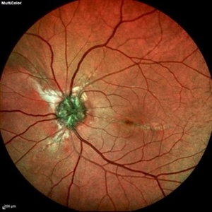 Optic Nerve Head Drusen With Angiod Streaks in Hyperphosphatemic Familial Tumoral Calcinosis
Aug 8 2024 by Hemanth Murthy, MBBS, MD, FASRS
Multicolor image of left eye of 53 year female patient with decreased vision in left eye. Patient gives history of multiple joint swellings with multiple dental procedures due to calcification of the roots. She had type2 MNV demonstrated on OCT and OCTA. Her blood reports showed elevated serum phosphorus (6.4 mg/dl) with normal serum calcium, vitamin D and parathyroid hormone. Her fibroblast growth factor 23 was markedly elevated(>1500RU/ml).
Photographer: Mr Veda Vyas
Condition/keywords: Optic disc drusen and Angiod streaks
-
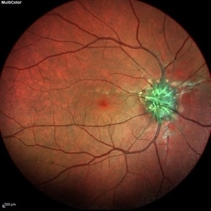 Optic Nerve Head Drusen With Angiod Streaks in Hyperphosphatemic Familial Tumoral Calcinosis
Aug 8 2024 by Hemanth Murthy, MBBS, MD, FASRS
Multicolor image of right eye of 53 year female patient with decreased vision in left eye. Patient gives history of multiple joint swellings with multiple dental procedures due to calcification of the roots. She showed type2 MNV on OCT and OCTA. Her blood reports showed elevated serum phosphorus (6.4 mg/dl) with normal serum calcium, vitamin D and parathyroid hormone. Her fibroblast growth factor 23 was markedly elevated(>1500RU/ml).
Photographer: Mr Veda Vyas
Condition/keywords: Optic disc drusen and Angiod streaks
-
 Optic Nerve Head Drusen With Angiod Streaks in Phosphatemic Familial Tumoral Calcinosis
Aug 8 2024 by Hemanth Murthy, MBBS, MD, FASRS
Autofluorescence image of left eye of 53 year female patient with decreased vision in left eye. Patient gives history of multiple joint swellings with multiple dental procedures due to calcification of the roots. She showed type 2 MNV in left eye on OCT and OCTA. Her blood reports showed elevated serum phosphorus (6.4 mg/dl) with normal serum calcium, vitamin D and parathyroid hormone. Her fibroblast growth factor 23 was markedly elevated(>1500RU/ml).
Photographer: Mr Veda Vyas
Condition/keywords: Optic disc drusen and Angiod streaks
-
 Optin Nerve Head Drusen With Angiod Streaks in Hyperphosphatemic Familial Tumoral Calcinosis
Aug 8 2024 by Hemanth Murthy, MBBS, MD, FASRS
Autofluorescence image of right eye of 53 year female patient with decreased vision in left eye. Patient gives history of multiple joint swellings with multiple dental procedures due to calcification of the roots. She had type2 MNV in left eye demonstrated on OCT and OCTA. Her blood reports showed elevated serum phosphorus (6.4 mg/dl) with normal serum calcium, vitamin D and parathyroid hormone. Her fibroblast growth factor 23 was markedly elevated(>1500RU/ml).
Photographer: Mr Veda Vyas
Condition/keywords: Optic disc drusen and Angiod streaks
-
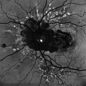
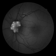
 Autofluorescence in Optic Nerve Head Drusen
May 28 2024 by Nishikant J Borse, MS, FMRF, FASRS
65-year-old female was referred for disc edema. An Autofluorescence Imaging was done which showed the autofluorescence of the optic nerve head drusen.
Photographer: Dr Nishikant Borse , Insight eye Clinic , Mumbai
Imaging device: Topcon Triton
Condition/keywords: Autofluorescence imaging of Optic Disc Drusen
-
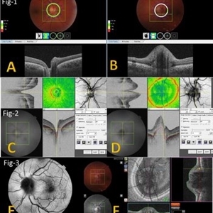 Multimodal Imaging for Differentiating Unilateral Pseudo Optic Disc Swelling(Buried Drusen) From True Optic Disc Swelling
Feb 7 2024 by Fawwaz F Al Mamoori, MD, Medical Retina Consultant
27-year-old male, medically free, presented with left unilateral optic disc swelling. BCVA=1.0(OU), color vision, and contrast sensitivity were normal (OU)with no RAPD in the left eye. Swept Source OCT: showed elevated left optic disc with hyporeflective mass (Fig-1 B). Enface OCT: Showed left peripapillary multiple ovoid mass lesions(drusen) (Fig-2 d, Fig3 F). FAF: of the left eye showed superonasal hyper autofluorescent drusenoid lesions)(Fig3 E). Orbital MRI with contrast was requested to exclude any compressive lesions like tumors(menigioma)or inflammatory lesions like granuloma(sarcoid granuloma). orbital MRI result was normal.
Photographer: Hana.S.Owais
Imaging device: TRITON(TOPCON,Swept Source OCT)
Condition/keywords: fundus autofluorescence (FAF), multimodal imaging, OCT EN FACE, optic disc drusen, optic disc edema, swept source
-
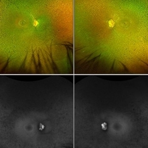
 Multimodal Imaging for Differentiating Unilateral Pseudo Optic Disc Swelling(Buried Drusen) From True Optic Disc Swelling
Feb 7 2024 by Fawwaz F Al Mamoori, MD, Medical Retina Consultant
A 27-year-old male patient, medically free, presented with unilateral left optic disc swelling. BCVA=1.0(OU), color vision, and contrast sensitivity were normal (OU) with no RAPD in the left eye. SS-OCT: showed left optic disc elevation with hyporeflective mass lesion (Fig-1 B). Enface OCT: showed left peripapillary hyperreflective ovoid mass lesions(Fig-2 D, Fig-3 F), FAF: showed left superonasal hyperautofluorescent drusenoid lesions. Orbital MRI with contrast was requested to exclude any optic nerve compressive lesions like (tumors: like mengioma or inflammatory lesions like granuloma (sarcoidosis). the result of orbital MRI was normal.
Photographer: Hana.S.Owais
Imaging device: TRITON(TOPCON,Swept Source OCT)
Condition/keywords: fundus autofluorescence (FAF), multimodal imaging, OCT EN FACE, optic disc drusen, optic disc edema
-
 Non invasive multimodal imaging for differentiating unilateral pseudo swelling buried optic disc drusen from true optic disc swelling
Feb 7 2024 by Fawwaz F Al Mamoori, MD, Medical Retina Consultant
27-year-old male, medically free, routine fundus examination showed left optic dic swelling, BCVA =1.0(OU), color vision, and contrast sensitivity were normal with no RAPD (OU). SS-OCT of the left optic disc showed a hyporeflective mass. Enface OCT shadogram showed peripapillary ovoid structures (drusen).FAF: showed drusenoid autofluorescence in the superonasal part only. Orbital MRI with contrast was requested to exclude any optic nerve tumor and it was normal.
Photographer: Hana.S.Owais
Imaging device: TRITON(OCT) Topcon
Condition/keywords: multimodal imaging, optic disc drusen, optic disc swelling
-
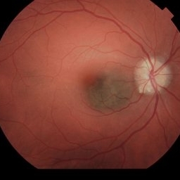 Suspicious Choroidal Nevus / Optic Disc Drusen
Nov 1 2023 by Virginia Gebhart
29 year-old female with suspicious choroidal nevus adjacent to optic nerve and extending into fovea. Optic disc drusen OU. Pt is asymptomatic
Photographer: Virginia Gebhart
Imaging device: Topcon
Condition/keywords: choroidal nevus, disc drusen, drusen of optic disc, nevus
-
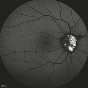
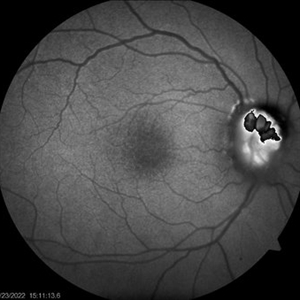
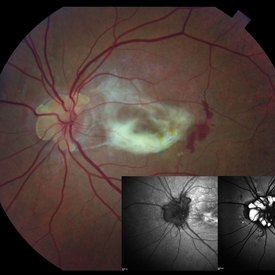
 Retinitis pigmentosa with disc drusen
Aug 6 2023 by AMIT NENE
Fundus photograph of a 42 year old female with advanced retinitis pigmentosa. Autofluorescence imaging showing associated optic disc drusen.
Photographer: Rahul Saradage, Isha Netralaya, Thane
Imaging device: Optos imaging
Condition/keywords: Autoflourescence, optic disc drusen, retinitis pigmentosa
-
 Optic Disc Drusen Autofluorescence
Jul 16 2023 by Aditya S Kelkar, MS, FRCS, FASRS,FRCOphth
Fundus autofluorescence imaging of a 22-year-old male with optic disc drusen seen as hyperautofluorescent spot.
Photographer: Dr. Harsh Jain
Condition/keywords: Autofluorescence imaging of Optic Disc Drusen
-
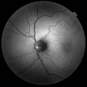
 Optic Disc Drusen
Jun 29 2022 by Mohamed Awadalla, MD, FRCSEd
Autofluorescence in optic disc drusen Red free fundus photo
Condition/keywords: Autofluorescence, optic disc drusen
-
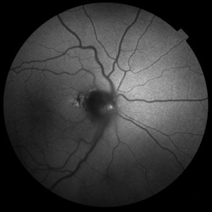
 ONH-drusen-OD
Mar 24 2022 by Elite Bor-Shavit, MD
Fundus autofluorescence of a 41-years old patient with combined true papilledema and optic nerve head drusen, treated with Diamox and monitored.
Condition/keywords: optic disc drusen, papilledema
-
 ONH-drusen-OS
Mar 24 2022 by Elite Bor-Shavit, MD
Fundus autofluorescence of a 41-years old patient with combined true papilledema and optic nerve head drusen, treated with Diamox and monitored.
Condition/keywords: optic disc drusen, papilledema
-
 Optic Disc Drusen and Angioid Streaks
Jun 3 2020 by Mirko Ratkovic, MD
Optic disc drusen and angioid streaks.
Condition/keywords: angioid streaks, fundus autofluorescence (FAF), optic disc drusen
-
 Optic Disc Drusen and Angioid Streaks
Jun 3 2020 by Mirko Ratkovic, MD
Optic disc drusen and angioid streaks.
Condition/keywords: angioid streaks, fundus autofluorescence (FAF), fundus photograph, optic disc drusen
-
 Optic Disc Drusen and Angioid Streaks
Jun 3 2020 by Mirko Ratkovic, MD
Optic disc drusen and angioid streaks.
Condition/keywords: angioid streaks, fundus autofluorescence (FAF), fundus photograph, optic disc drusen
-
 Optic Disc Drusen and Angioid Streaks
Jun 3 2020 by Mirko Ratkovic, MD
Optic disc drusen and angioid streaks.
Condition/keywords: angioid streaks, drusen of optic disc, fundus autofluorescence (FAF), fundus photograph
-
 Angioid Streaks with Associated Disc Drusen and CNV
Jan 6 2020 by Sarah Oelrich
Angioid streaks with associated disc drusen and CNV.
Photographer: Sarah Oelrich CRA COT OCT-C
Imaging device: Topcon, Heidelberg
Condition/keywords: angioid streaks, choroidal neovascular membrane (CNVM), disc drusen
 Loading…