 Initializing download.
-
 By Hemanth Murthy, MBBS, MD, FASRS
RETINA INSTITUTE OF KARNATAKA
Co-author(s): Dr Sumanth A M, Dr Prakruthi - Uploaded on Aug 8, 2024.
- Last modified by Joshua Friedman on Aug 9, 2024.
- Rating
- Appears in
- 8-Aug-2024
- Condition/keywords
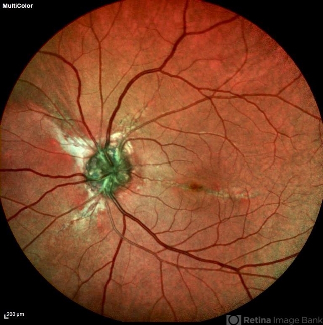
- Optic disc drusen and Angiod streaks
- Photographer
- Mr Veda Vyas
- Imaging device
- Scanning laser ophthalmoscope
- Description
- Multicolor image of left eye of 53 year female patient with decreased vision in left eye. Patient gives history of multiple joint swellings with multiple dental procedures due to calcification of the roots. She had type2 MNV demonstrated on OCT and OCTA. Her blood reports showed elevated serum phosphorus (6.4 mg/dl) with normal serum calcium, vitamin D and parathyroid hormone. Her fibroblast growth factor 23 was markedly elevated(>1500RU/ml).


