Search results (419 results)
-
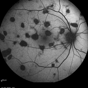
 Toxoplasma Retinochoroiditis
Dec 21 2025 by Gyanendra Lamichhane
24 female, Focal Retinitis, posterior pole involvement with focal vitiritis,
Photographer: Prof.Gyanendra Lamichhane
Condition/keywords: posterior uveitis, Retinochoroiditis, Toxoplasma
-
 LE Serpiginous Like Choroiditis
Dec 21 2025 by Gyanendra Lamichhane
25 year-old male, presented with decreased vision LE 2 weeks, note Serpigious like involvement without not involving Disc. This is quite common in TB endemic areas.
Photographer: Prof.Dr Gyanendra Lamichhane
Condition/keywords: serpiginous like choroiditis
-
 Chorioretinitis
Dec 16 2025 by Kimberly Wakester
Optomap RGB of a 37 year-old woman with Chorioretinitis. The chorioretinitis remains largely stable in both eyes on exam and compared to prior photos. Clinical and diagnostic findings in both eyes continue to be most consistent with punctate inner choroidopathy (PIC) /Multifocal choroiditis and panuveitis (MCP). Will continue follow up care every 6 months with dilated exam and repeat testing.
Photographer: Kimberly Wakester, COA, OCT-C
Imaging device: Optos California
Condition/keywords: chorioretinitis, multifocal chorioretinitis (MCP), punctate inner choroidopathy (PIC)
-
 Multifocal Choroiditis with Panuveitis
Oct 16 2025 by Virginia Gebhart
39 year old female diagnosed with MCP in 2009. Extensive RPE changes and hypertrophy, arterial attenuation and pale nerve. Currently no active inflammation.
Photographer: Virginia Gebhart, Retina Consultants of Carolina
Imaging device: Optos California
Condition/keywords: corticosteroid-induced glaucoma, hypertrophy, multifocal chorioretinitis (MCP), PALE DISC
-
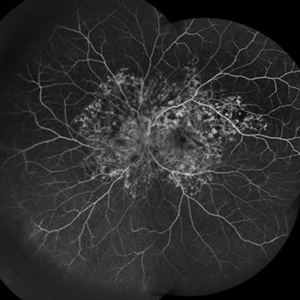 Snaking Away
Sep 1 2025 by Malvika Singh
Fluorescein angiography montage of a 45 year old man showing areas of staining in a case of healed choroiditis.
Photographer: Dr Malvika Singh, Retina Foundation, Ahmedabad, India
Imaging device: Mirante SLO/OCT
Condition/keywords: fluorescein angiogram (FA), healed choroiditis, serpiginous choroiditis
-
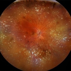
 Healed AZOOR- Multiple White Dot Syndrome
Aug 29 2025 by Aditya S Kelkar, MS, FRCS, FASRS,FRCOphth
Fundus photograph of a 58 year old woman with multiple, well-defined, punched-out chorioretinal scars scattered throughout the posterior pole and mid-periphery. The macular area shows a large, confluent, yellowish-white scar involving the fovea.
Photographer: Dr. Muskan Mangal
Imaging device: Optos Daytona
Condition/keywords: multifocal choroiditis, multiple evanescent white dot syndrome (MEWDS), punctate inner choroidopathy (PIC)
-
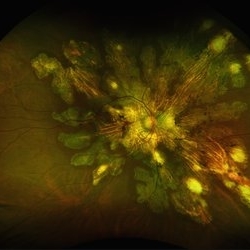
 Ghost Map Retina
Aug 4 2025 by Malvika Singh
Fundus photograph of a 50 year old male showing extensive chorioretinal scarring.
Photographer: Dr Malvika Singh, Retina Foundation, Ahmedabad, India
Imaging device: Mirante SLO/OCT
Condition/keywords: healed choroiditis
-
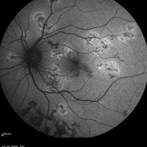
 Serpiginous Choroidopathy
Jun 23 2025 by César Adrián Gómez Valdivia, MD
Fundus photograph of a 29 year-old female patient diagnosed with Serpiginous Choroidopathy. Finings were bilateral. The most common complication of SC is choroidal neovascularization affecting up to 35% of patients. Other reported complications are subretinal fibrosis, cystoid macular edema, branch vein occlusion, serous retinal detachment, optic disc neovascularization ,and anterior uveitis.
Photographer: @eyemissu2
Imaging device: TOPCON TRC-50DX
Condition/keywords: serpiginous choroiditis
-
 Serpiginous Choroidopathy
Jun 23 2025 by César Adrián Gómez Valdivia, MD
Fundus photograph of a 29 year-old female patient diagnosed with Serpiginous Choroidopathy. Finings were bilateral. The most common complication of SC is choroidal neovascularization affecting up to 35% of patients. Other reported complications are subretinal fibrosis, cystoid macular edema, branch vein occlusion, serous retinal detachment, optic disc neovascularization, and anterior uveitis.
Photographer: @eyemissu2
Imaging device: California ICG OPTOS
Condition/keywords: serpiginous choroiditis
-
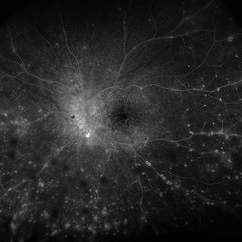
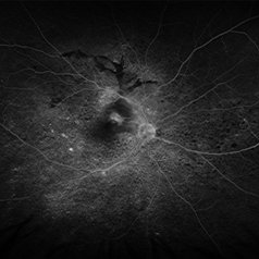
 Active Multi Focal Choroiditis
Jun 21 2025 by Moazzam Parvez
Auto fluorescence image of a 28 year old gentleman with active multifocal choroiditis in his left eye and healed choroiditic patches in the right eye.
Photographer: Moazzam Parvez , Netralayam , Kolkata
Imaging device: Heidelberg Spectralis
Condition/keywords: active, multifocal choroiditis
-
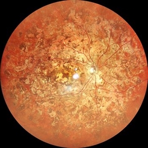
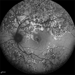
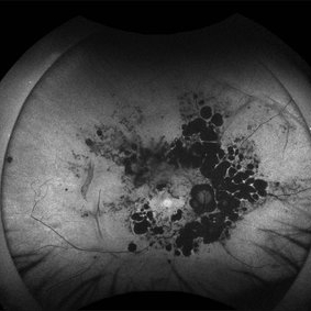
 Fractal Pattern of Chronic Serpiginous Choroiditis
Jun 17 2025 by Guilherme Sturzeneker, MD, MSc
Ultra-widefield fundus photograph and autofluorescence of a 33-year-old woman with longstanding serpiginous choroiditis in the right eye. The image reveals centrifugal chorioretinal atrophy forming a dramatic fractal-like pattern, sparing the fovea. The patient is several years post-onset, with repeated negative workups, including for tuberculosis. Despite extensive lesions, the patient retains 20/20 vision in both eyes. Management included azathioprine monotherapy, as systemic steroids were contraindicated due to bipolar disorder.
Photographer: Andrea Almeida, IPEPO - Instituto da Visão
Imaging device: Optos Silverstone
Condition/keywords: autoimmune uveitis, azathioprine, chorioretinal atrophy, serpiginous choroiditis, ultra-wide field imaging
-
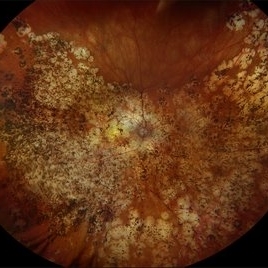
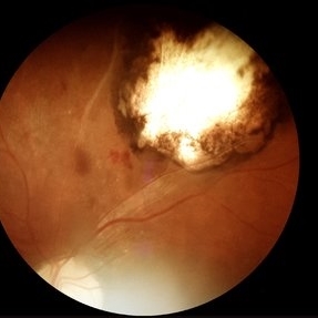
 The Headlight in the Fog
Jun 17 2025 by Thirumalesh Mochi Basavaraj, MD
37 year old male with sudden onset diminution of visual acuity has a large retinochoridal granuloma along the superotemporal arcade and a few with satellite lesions more temporal to it, there was extensive Occlusoive vasculitis (both arterioles and veins )being involved with Vitrities.
Photographer: Vivekanand ,Narayana nethralaya
Imaging device: Daytona
Condition/keywords: acute toxoplasmosis, retinochoroiditis
-
 Idiopathic Multifocal Choroiditis
Jun 14 2025 by César Adrián Gómez Valdivia, MD
Fundus Autofluorescence of an 46 YO female patient diagnosed with Idiopathic Multifocal Choroiditis. Findings were bilateral.
Photographer: @eyemissu2
Imaging device: California ICG OPTOS
Condition/keywords: choroiditis
-
 VKH Syndrome
Jun 12 2025 by Virginia Gebhart
22 year old male with VKH Syndrome. Pt has been experiencing severe headaches, distorted vision, hearing loss, weakness, and a large white patch of hair. Significant cell in AC and vitreous, multiple punched-out CR scars in periphery. Referred to rheumatology for possible immunomodulatory treatment
Photographer: Virginia Gebhart, Retina Consultants of Carolina
Imaging device: Optos California
Condition/keywords: montage, multifocal choroiditis, panuveitis, Vogt-Koyanagi-Harada
-
 VKH Syndrome
Jun 12 2025 by Virginia Gebhart
Fluorescein angiogram of 22 year old male with VKH syndrome. Significant cell in AC and vitreous, multiple punched-out CR scars in periphery, mild vascular leakage. Pt referred to rheumatology for immunomodulatory treatment.
Photographer: Virginia Gebhart, Retina Consultants of Carolina
Imaging device: Optos California
Condition/keywords: FA, fluorescein angiogram (FA), multifocal choroiditis, panuveitis, VKH, Vogt-Koyanagi-Harada
-
 Active multifocal choroiditis
May 26 2025 by Moazzam Parvez
Auto fluorescence photograph of an 43 year old man with active choroiditic lesion present in the left eye with recurrence
Photographer: Dr Moazzam Parvez , Netralayam , Kolkata
Imaging device: Heidelberg Spectralis
Condition/keywords: active choroididtis, choroiditi
-
 Healed Choroiditis
May 14 2025 by Moazzam Parvez
Auto fluorescence image of a 42 year old woman with healed choroiditic patches.
Photographer: Dr Moazzam Parvez, Netralayam, Kolkata
Imaging device: Heidelberg Spektrales
Condition/keywords: multifocal choroiditis
-
 VKH Pseudotumor – Chronic Subretinal Fibrosis
May 11 2025 by Felipe Murati
Ultra-widefield fundus image from a 36-year-old woman with chronic VKH syndrome showing a pseudotumor-like subretinal fibrotic lesion in the right eye. The lesion developed after multiple relapses and remained stable over a 1-year follow-up with immunosuppressive treatment including prednisone, mycophenolate mofetil, and adalimumab. No active choroiditis or exudative detachment was observed. Multimodal imaging was essential for disease monitoring.
Photographer: Felipe A. Murati, MD, University of Arizona
Imaging device: Optos California ultra-widefield retinal imaging system, single-capture, color fundus modality.
Condition/keywords: adalimumab, chronic inflammation, granulomatous uveitis, OCT, Optos ultra-widefield imaging, pseudotumor, subretinal fibrosis, VKH, Vogt-Koyanagi-Harada
-
 VKH Pseudotumor – Fluorescein Angiography
May 11 2025 by Felipe Murati
Fluorescein angiography image from a 36-year-old woman with chronic Vogt-Koyanagi-Harada (VKH) syndrome showing a pseudotumor-like lesion with late-phase staining and no active leakage. The image highlights subretinal fibrosis in the right eye, stable under long-term immunosuppressive therapy with mycophenolate mofetil and adalimumab. No signs of active choroiditis are present, confirming a quiescent phase.
Photographer: Felipe A. Murati, MD, University of Arizona
Imaging device: Optos California, fluorescein angiography modality
Condition/keywords: choroiditis, Fluorescein angiography, granulomatous uveitis, Optos FA, pseudotumor, subretinal fibrosis, VKH, Vogt-Koyanagi-Harada
-
 Retinocoroiditis Inactiva Por Toxoplasmosis
Apr 28 2025 by Paulina Araujo
Fundus photography demonstrates a 2-disc-diameter chorioretinal scar in the superior temporal arcade, consistent with inactive toxoplasmic retinochoroiditis. The lesion exhibits pigmented borders and central atrophy, with adjacent splinter hemorrhages and vascular sheathing. No vitreous inflammation or active satellite lesions are present.
Photographer: Paulina D.Araujo Martínez, Asociación para Evitar la Ceguera en México I.A.P., Hospital Dr Luis Sánchez Bulnes.
Condition/keywords: toxoplasmosis chorioretinitis
-
 Serpiginous Choroidopathy
Oct 19 2024 by César Adrián Gómez Valdivia, MD
Fundus photograph of a 29-year-old woman with Serpiginous Choroidopathy. Finings were bilateral.
Photographer: @eyemissu2
Imaging device: California ICG OPTOS
Condition/keywords: macula serpiginous choroidopathy, serpiginous choroiditis, serpiginous like choroiditis
-
 Ocular Toxoplasmosis
Sep 24 2024 by Gustavo Uriel Fonseca Aguirre
24-year-old patient with a history of retinochoroiditis due to toxoplasmosis in the right eye, a focus of retinochoroiditis reactivation of toxoplasmosis is observed.
Photographer: Gustavo U. Fonseca Aguirre, Fundación Hospital Nuestra Señora de la Luz, Ciudad de México
Condition/keywords: Kyrieleis arteritis, toxoplasmosis reactivation
-
 Serpiginous Choroidopathy Autofluorescence
Sep 24 2024 by Gustavo Uriel Fonseca Aguirre
Autofluorescence image of the right fundus of a 32-year-old female patient diagnosed with serpiginous choroiditis.
Photographer: Gustavo U. Fonseca Aguirre, Fundación Hospital Nuestra Señora de la Luz, Ciudad de México
Condition/keywords: autofluorescence imaging, serpiginous choroiditis
-
 Serpiginous Choroidopathy
Sep 24 2024 by Gustavo Uriel Fonseca Aguirre
Right fundus of a 32-year-old female patient diagnosed with serpiginous choroiditis.
Photographer: Gustavo U. Fonseca Aguirre, Fundación Hospital Nuestra Señora de la Luz, Ciudad de México
Condition/keywords: Fundus examination, serpiginous choroiditis
 Loading…