Search results (398 results)
-
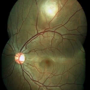
 Disseminated Retinitis and Retinochoroiditis, Metastatic
May 16 2017 by Karen Panzegrau
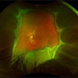
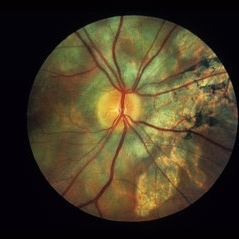
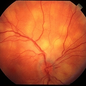
Fundus photograph of 44-year-old male with plasmacytoma infiltation of the choroid confirmed by biopsy, associated with disseminated retinitis, and retinochoroiditis. Vision is LP. Patient treated with intravitreal methotrexate
Photographer: Karen Panzegrau
Imaging device: Optos
Condition/keywords: metastatic lesion, methotrexate, Optos, plasmacytoma, retinitis, retinochoroiditis, unilateral exudative retinal detachment
-
 Tuberculosis-related serpiginous-like choroiditis
Nov 22 2022 by Ricardo Leitão Guerra
True color BLFI of a 60-year-old male presenting chorioretinal scars from a tuberculosis-related serpiginous-like choroiditis.
Photographer: Ricardo Leitão Guerra
Imaging device: Zeiss Clarus 700
Condition/keywords: serpiginous choroiditis, tuberculosis
-
 Serpiginous Choroiditis
Sep 22 2019 by Haider Ali
35-year-old female presented with decrease in vision in her left eye for last 4 days and in right eye for last 8 days. Her right eye was previously involved in a similar episode about 5-6 months ago for which she was treated with oral steroids.
Photographer: Dr Haider Ali Chaudhry, Madinah Teaching Hospital, Faisalabad
Condition/keywords: acute posterior multifocal placoid pigment epitheliopathy (APMPPE), macula serpiginous choroidopathy, posterior uveitis, serpiginous choroiditis, uveitis, white dot lesions, white dot syndrome
-
 Toxoplasma Retinochoroiditis
Feb 25 2013 by Henry J. Kaplan, MD
Toxoplasmosis, right eye: reactivation of congenital toxoplasmosis as an active retinitis lesion with overlying vitritis adjacent to an old scar.
Condition/keywords: toxoplasmosis chorioretinitis, toxoplasmosis reactivation
-
 Macular Serpiginous Choroidopathy
Sep 27 2012 by Raj K. Maturi, MD
9/11/2012
Photographer: Char Harris
Imaging device: HRA
Condition/keywords: serpiginous choroiditis
-
---thumb.jpg/image-square;max$300,300.ImageHandler) Multifocal Choroiditis
Multifocal Choroiditis
Feb 26 2013 by Henry J. Kaplan, MD
Multifocal choroiditis, MFC, inactive scars in the periphery.
Condition/keywords: multifocal choroiditis
-
 Multifocal Choroiditis
Jul 19 2020 by Aditya S Kelkar, MS, FRCS, FASRS,FRCOphth
Fundus photograph of 29-year-old male with both eyes inactive multifocal choroiditis.
Photographer: Dr. Sayali Tidke
Imaging device: CLARUS 500
Condition/keywords: multifocal choroiditis
-
 Multifocal Choroiditis
Aug 16 2018 by FELIPE PEREIRA
Mid-phase indocyanine green angiography of a 25-year-old woman with sudden central vision loss and photopsias for 7 days. The hypofluorescent lesions in the macula and nasal to the disc correspond to the yellow-white deep lesions in the fundus examination. No leakage is observed at any stage of the exam
Photographer: Claudio Zett Lobos
Imaging device: HEIDELBERG SPECTRALIS HRA
Condition/keywords: indocyanine green (ICG) angiography, multifocal choroiditis, white dot syndrome
-
 Serpiginous Choroiditis
Jun 4 2014 by Henry J. Kaplan, MD
The same patients more nasally demonstrates scar formation and outer retinal atrophy. #2
Condition/keywords: serpiginous choroiditis
-
 Serpiginous Choroiditis
Sep 22 2019 by Haider Ali
35-year-old female presented with decrease in vision in her left eye for last 4 days and in right eye for last 8 days. Her right eye was previously involved in a similar episode about 5-6 months ago for which she was treated with oral steroids.
Photographer: Dr Haider Ali Chaudhry, Madinah Teaching Hospital, Faisalabad
Condition/keywords: acute posterior multifocal placoid pigment epitheliopathy (APMPPE), macula serpiginous choroidopathy, posterior uveitis, serpiginous choroiditis, uveitis, white dot lesions, white dot syndrome
-
 Serpiginous Choroiditis
Nov 14 2021 by Maxwell J Wingelaar, MD
An image showing active Serpiginous Choroiditis.
Condition/keywords: serpiginous choroiditis
-
 Serpiginous Choroiditis (Recurrent)
Sep 22 2019 by Haider Ali
35-year-old female presented with decrease in vision in her left eye for last 4 days and in right eye for last 8 days. Her right eye was previously involved in a similar episode about 5-6 months ago for which she was treated with oral steroids.
Photographer: Dr Haider Ali Chaudhry, Madinah Teaching Hospital, Faisalabad
Condition/keywords: acute posterior multifocal placoid pigment epitheliopathy (APMPPE), macula serpiginous choroidopathy, serpiginous choroiditis, white dot syndrome
-
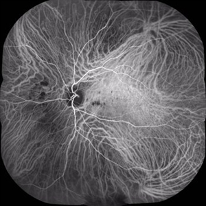
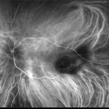
 Serpiginous Choroiditis - Fundus Autofluorescence
Sep 20 2014 by Rameez N Hussain, MD
Fundus autofluorescence of serpiginous choroiditis showing decreased autofluorescence area corresponding to the inactive lesion (RPE atrophy) and increased autofluorescence area corresponding to active lesion.
Photographer: Dr.Rameez N Hussain, MD, Central Imaging Center, Vitreo Retinal Services, Giridhar Eye Institute, Cochin, India
Imaging device: Heidelberg Blue Peak Autofluorescence imaging.
Condition/keywords: serpiginous choroiditis
-
 Serpiginous Choroiditis With Peripapillary SRNVM
Apr 17 2017 by Manish Nagpal, MD, FRCS (UK), FASRS
patient having serpiginous choroiditis came with recent drop of central vision. Fundus revealed a streak of blood lining a membrane in the peripapillary lesion suggestive of a SRNVM.
Photographer: pooja barot
Condition/keywords: choroiditis, serpiginous choroiditis, subretinal neovascular membrane
-
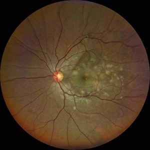 Tubercular Multifocal Choroiditis
Aug 18 2021 by Priyanka Raj, MBBS, MS
Fundus photograph of a 28 year-old man with multifocal tubercular choroiditis.
Photographer: Priyanka Raj, Prakash Netra Kendr, Lucknow, India
Imaging device: Zeiss Clarus 500
Condition/keywords: choroiditis, tuberculosis
-
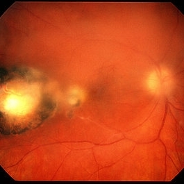
 West Nile Virus Choroiditis
Apr 4 2014 by Suber S. Huang, MD, MBA, FASRS
Fundus photograph (11 month follow-up) of an 88-year-old woman who developed West Nile virus encephalitis on 08/2012 and subsequent choroiditis.
Photographer: Geoffrey Pankhurst; University Hospitals Eye Institute, Case Western Reserve University, Cleveland, OH
Imaging device: TopCon TRC50EX
Condition/keywords: choroiditis, disseminated choroiditis, infectious uveitis, optic nerve atrophy
-
---thumb.jpg/image-square;max$300,300.ImageHandler) Multifocal Choroiditis and Panuveitis Syndrome
Multifocal Choroiditis and Panuveitis Syndrome
Feb 26 2013 by Henry J. Kaplan, MD
Multifocal choroiditis and panuveitis: left eye. Acute stage: haziness of the media due to vitritis and multiple active yellow and also inactive choroidal lesions are present.
Condition/keywords: multifocal choroiditis
-
 Sarcoidosis Choroiditis
Feb 25 2013 by Henry J. Kaplan, MD
Sarcoidosis multifocal choroiditis in a case with a known diagnosis of sarcoidosis.
Condition/keywords: sarcoidosis choroiditis
-
 Serpiginous Choroiditis
Feb 25 2013 by Henry J. Kaplan, MD
Typical serpiginous choroiditis: right eye.
Condition/keywords: serpiginous choroiditis
-
 Serpigenous Choroidopathy in a 68-Year-Old Male
Feb 15 2013 by Roy Schwartz, MD
A 68-year-old healthy male presented with a few years of decreased vision bilaterally. Visual acuity in OD was 1/36 and in OS 20/40. Anterior segments were normal except for bilateral mild nuclear sclerosis and pseudoexfoliation in OS. In the fundus of OD a large atrophy with pigmentary scars were seen in the macula and nasally to the optic disc while OS presented with the same clinical picture but an island of normal appearing retina was seen in the fovea. On fluorscein angiography no leakage was shown. A diagnosis of Serpigenous choroidopathy was made.
Photographer: Galit Yair-Pur
Condition/keywords: macula serpiginous choroidopathy, serpiginous choroiditis
-
 ICG: Choroidal Aspergilloma With Secondary Choroidal Neovascularization and Exudative Retinal Detachment
Mar 21 2019 by Scott D Walter, MD, MSc, FASRS
Multimodal imaging of a transplant patient with disseminated Aspergillosis and vision loss in her left eye.
Condition/keywords: choroidal neovascular membrane (CNVM), choroidal neovascularization (CNV), exudative detachment, focal chorioretinitis, fungal endophthalmitis, granulomatous choroiditis
-
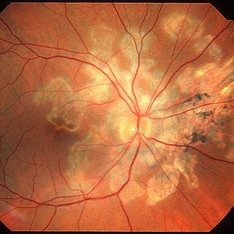
 Serpiginous Choroiditis
Feb 25 2013 by Henry J. Kaplan, MD
Serpiginous choroiditis, left eye. Active yellowish edematous lesion visible around the optic nerve toward the fovea and also old pigmented scar in the fovea.
Condition/keywords: serpiginous choroiditis
-
 Choroidal Granuloma
Apr 23 2019 by Purva Patwari
22-year-old male patient presented with blurring of vision in the right eye noticed since last one week. He was asymptomatic a week ago when he noticed the blurring in his right eye. On examination his vision was 6/6 in both eyes. Anterior segment was normal. Posterior segment was normal for the left eye. Right eye examination revealed a clear vitreous cavity with choroidal granulomas scattered throughout the fundus. The present picture shows choroidal granulomas with OCT segment passing through the parafoveal lesion showing subretinal fluid accumulation and corresponding thickening of the retinal layers. CT scan reveals heterogeneously enhancing lymph nodes showing conglomerationin the hilar region-possibility of tubercular etiology.
Photographer: Dr Purva Patwari, Patwari Retina Center
Imaging device: Zeiss Visu 500
Condition/keywords: choroidal granuloma, choroiditis, granulomatous choroiditis, tubercular choroidal granuloma, tuberculosis
-
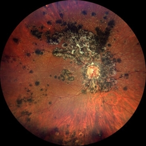
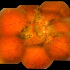
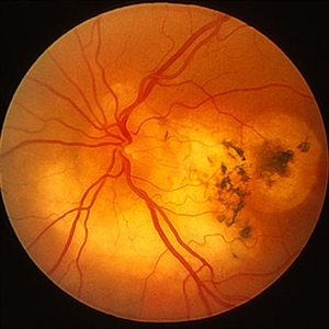
 Peripheral Choroidal Granuloma Associated With Tuberculosis Choroiditis
Jun 3 2017 by S. Natarajan, MD, FASRS, FRCS (GLASGOW) , FICO, D.Sc, FELA
Funds photograoh of an 21-year-old female pheripheral choroidal granuloma associated with tuberculos choroiditis.
Photographer: miss ashwini borde
Imaging device: Carl Zeiss 450 Plus IR
Condition/keywords: peripapillary choroidal granuloma
-
 Serpiginous
Aug 29 2012 by F. Ryan Prall, MD
70-year-old female with reactivation of serpiginous choroiditis.
Photographer: Tom Egnatz, Indiana University
Condition/keywords: serpiginous choroiditis
 Loading…